Background
Contribution of Bremsstrahlung X-rays to the X-ray counts under a characteristic elemental X-ray peak.
Backscatter
When X-rays interact with low average atomic number samples (for example, polyethylene or other low density materials), X-rays can be scattered back toward the instrument's detector and observed as part of the background component of a spectra.
Backscattered electrons
Come from the top half of the interaction volume (upper 400 nm) and provide atomic (Z) number information.
Beam current/probe current/spot size
Reflects the flow of electrons in the primary beam of the electron microscope. The magnitude of electron flow in the electron beam is directly proportional to the number of X-rays generated from the sample and the number of X-ray counts (intensities) recorded in the X-ray spectrum. Increasing the beam current will increase the number of X-rays generated from the sample but will not change the relative heights (intensities) of the characteristic X-ray peaks in the spectrum.
Binarization
This Is the process of turning a color or grayscale image into a binary (B&W) image.
Binary image
A binary image is a digital image that has only two possible values for each pixel. Typically, the two colors used for a binary image are black and white.
Bremsstrahlung
Electromagnetic radiation produced by the deceleration of a charged particle when deflected by another charged particle, typically an electron by an atomic nucleus. The term is also used to refer to the process of producing the radiation. Bremsstrahlung has a continuous spectrum, which becomes more intense and whose peak intensity shifts toward higher frequencies as the change of the energy of the decelerated particles increases.
Certified Reference Material (CRM)
These are 'controls' or standards used to check the quality and metrological traceability of products, to validate analytical measurement methods, or for the calibration of instruments. A certified reference material is a particular form of measurement standard. Reference materials are particularly important for analytical chemistry and clinical analysis. Since most analytical instrumentation is comparative, it requires a sample of known composition (reference material) for accurate calibration.
Characteristic X-rays
These X-rays are emitted when outer-shell electrons fill a vacancy in the inner shell of an atom, releasing X-rays in a pattern that is "characteristic" to each element. Characteristic X-rays were discovered by Charles Glover Barkla in 1909, who later won the Nobel Prize in Physics for his discovery in 1917.
Compton scatter
Compton inelastic scattering, discovered by Arthur Holly Compton, is the scattering of a photon after an interaction with a charged particle, usually an electron. If it results in a decrease in energy (increase in wavelength) of the photon (which may be an X-ray or gamma ray photon), it is called the Compton effect. Part of the energy of the photon is transferred to the recoiling electron.
Continuum
In physics, a continuous spectrum usually means a set of attainable values for some physical quantity (such as energy or wavelength) that is best described as an interval of real numbers, as opposed to a discrete spectrum, a set of attainable values that is discrete in the mathematical sense, where there is a positive gap between each value and the next one.
Detector window
For X-ray fluorescence spectroscopy, radiolucent windows are needed to protect X-ray sensors and to hold a vacuum. These windows can work as a transparent gas barrier for X-ray tubes and detectors. Because low-Z elements' characteristic spectra occur at low energies, it is necessary that these thin windows are made of low atomic number materials.
Duane-Hunt limit
The Duane–Hunt law, named after the American physicists William Duane and Franklin L. Hunt, gives the maximum frequency of X-rays that can be emitted by Bremsstrahlung in an X-ray tube by accelerating electrons through an excitation voltage into a metal target.
Dwell time
The time spent collecting X-rays for each pixel in an elemental map. EDS minimum map dwell is 50us while maximum is 100s. For Atlas, the minimum dwell is 1ms while maximum is 100s.
EDXRF
Energy Dispersive X-ray Fluorescence (EDXRF) is one of two general types of X-ray Fluorescence techniques used for elemental analysis applications. In EDXRF spectrometers, all of the elements in the sample are excited simultaneously, and an energy dispersive detector in combination with a multi-channel analyzer is used to simultaneously collect the fluorescence radiation emitted from the sample and then separate the different energies of the characteristic radiation from each of the different sample elements. Resolution of EDXRF systems is dependent upon the detector, and typically ranges from 125 eV (SDD) – 600 eV (proportional counter). The principal advantages of EDXRF systems are their simplicity, fast operation, lack of moving parts, and high source efficiency.
Electron configuration
In atomic physics and quantum chemistry, the electron configuration is the distribution of electrons of an atom or molecule (or other physical structure) in atomic or molecular orbitals. For example, the electron configuration of the neon atom is 1s2 2s2 2p6, meaning that the 1s, 2s and 2p subshells are occupied by 2, 2 and 6 electrons respectively. Electronic configurations describe each electron as moving independently in an orbital, in an average field created by all other orbitals. Mathematically, configurations are described by Slater determinants or configuration state functions. According to the laws of quantum mechanics, for systems with only one electron, a level of energy is associated with each electron configuration and in certain conditions, electrons are able to move from one configuration to another by the emission or absorption of a quantum of energy, in the form of a photon.
Electron trap
An attachment on the tip of a silicon drift detector (SDD) that prevents scattered electrons, from the SEM, from entering the detector and causing damage.
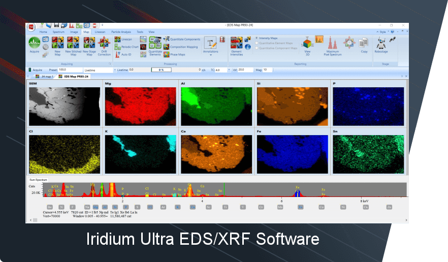
Elemental map
Elemental mapping is based on compiling extremely specific elemental composition data across an area of a sample. This is typically done in an SEM or TEM using EDS analysis. A high resolution image of the area of interest is collected along with the EDS data, and the two are correlated.
Energy calibration
XRF spectra are typically measured with MCAs (Multi Channel Analyzers) which holds an array of intensities. An XRF spectrum can be represented well with an MCA trace (intensities per bin) as long as one can convert bin number to energy. Fortunately, most measurement systems in use have a linear relation (calibration) between bin number and energy, and the use of an MCA trace as an XRF spectrum is straightforward.
Escape peaks
Escape peaks arise when a strong element peak is recorded. Their formation occurs within the detector crystal. When an incident photon is passing through the detector volume and its characteristic energy is sufficiently high, it can produce a photoelectron from an inner shell of a crystal atom (Si). As a result, the excited atom can emit a fluorescence X-ray photon, mostly a Kα photon. It is most of the times reabsorbed thus contributing to the charge pulse. However, that photon can also escape from the crystal. In that case, it carries off the definite energy of the Si− Kα. The charge pulse appear as corresponding to a photon energy Einitial − ESi − Kα and therefore show up in the spectrum as separate spurious peaks.
Fundamental Parameters (FP)
The FP method is a first-principles calculation method of chemical element concentration from the measured XRF spectra using the FPs such as the X-ray absorption coefficients, fluorescence yields, jump ratios, branching ratios, and the incident spectrum from the X-ray tube.
Full Width Half Max (FWHM)
In a distribution, full width at half maximum (FWHM) is the difference between the two values of the independent variable at which the dependent variable is equal to half of its maximum value. In other words, it is the width of a spectrum curve measured between those points on the y-axis which are half the maximum amplitude. This value determines the resolution of the peak, and under proper settings, the resolution capabilities of the detector. This value is reported in eV.
Gaussian deconvolution
Software subroutine that contains the parameters used to setup the various Gaussian deconvolution options used to extract net peak intensities from an spectrum with overlapped peaks. There are two options for the Gaussian method, which are the linear or nonlinear deconvolution. Non-linear deconvolution to automatically computes the Gain Factor, Offset, and FWHM.
Instrument detection limit (IDL)
The best possible limit of detection, calculated from a “clean” sample, i.e., one with no interferences.
Intensity
Since the XRF signal intensity for each atomic species correlates directly to the number of atoms present, the XRF signal can be used as a direct measurement of the composition and/or thickness of materials. Intensity is the number of X-rays produced per unit time, which is typically reported as counts per second (cps).
Ionization energy
In physics and chemistry, ionization energy (IE) is the minimum energy required to remove the most loosely bound electron of an isolated neutral gaseous atom or molecule. It is quantitatively expressed as X(g) + energy ⟶ X+(g) + e− where X is any atom or molecule, X+ is the resultant ion when the original atom was stripped of a single electron, and e− is the removed electron. Ionization energy is positive for neutral atoms, meaning that the ionization is an endothermic process. Roughly speaking, the closer the outermost electrons are to the nucleus of the atom, the higher the atom's ionization energy. In physics, ionization energy is usually expressed in electronvolts (eV) or joules (J). In chemistry, it is expressed as the energy to ionize a mole of atoms or molecules, usually as kilojoules per mole (kJ/mol) or kilocalories per mole (kcal/mol).
K-alpha (Kα)
Characteristic X-rays are produced when an element is bombarded with high-energy particles (such as photons). When the incident particle strikes a bound electron in an atom, a target electron is ejected from the inner shell of the atom. After the electron has been ejected, the atom is left with a vacant energy level, also known as a core hole. Outer-shell electrons then fall into the inner shell, emitting quantized photons with an energy level equivalent to the energy difference between the higher and lower states. K-alpha emission lines result when an electron transitions to a vacancy in the innermost "K" shell (principal quantum number n = 1) from a p orbital of the second, "L" shell (n = 2), leaving a vacancy there.
Least squares (LS)
The method of least squares is a standard approach in regression analysis to approximate the solution of overdetermined systems (sets of equations in which there are more equations than unknowns) by minimizing the sum of the squares of the residuals (a residual being: the difference between an observed value, and the fitted value provided by a model) made in the results of each individual equation. The most important application is in data fitting. The best fit in the least-squares sense minimizes the sum of squared residuals.
Limit of detection (LOD or LoD)
The limit of detection LOD (or detection limit, DL) is the lowest possible concentration at which the method can detect (but not quantify) the analyte within the matrix with certain degree of confidence. It is also defined as the lowest concentration that can be separated from a background noise with some reliability. In the analytical sciences, the so-called three-sigma rule of thumb (or 3σ rule) expresses a conventional heuristic that nearly all values are taken to lie within three standard deviations of the mean, and thus it is empirically useful to treat 99.7% probability as near certainty.
Line series
Characteristic X-ray emission spectra consist of spectral series (K, L, M, N…), whose lines have a common initial state with the vacancy in the inner level. All electron levels with the principal quantum number n equal to 1, 2, 3, 4, etc. are named as K, L, M, N etc. levels and denoted with corresponding Greek letters and digit indexes. The dependence of X-ray emission line energy on atomic number Z is defined by Moseley's law.
Livetime
A measure of the period of time in which the system is able to accept another signal for processing. Livetime + Deadtime = Total Acquisition (real) Time. Causes of Deadtime include: peak pile-up rejection circuitry, the FET reset process, the signal conversion process, and the ADC/MCA analysis process time.
Major elements
Major elements are generally considered to be elements that constitute more than 1% of a sample by weight.
Map Resolution
The number of pixels present in an elemental map regardless of the map size. Map resolution is not the same as spatial resolution.
Match
Analysis of a spectrum by comparing it with stored known sample spectra. Best matching spectra are typically displayed along with a goodness of fit metric. This feature can deliver either qualitative or rigorously quantitative results (using FP or LS).
Map Resolution
The number of pixels present in an elemental map regardless of the map size. Map resolution is not the same as spatial resolution.
MultiChannel Analyzer (MCA)
A multichannel analyzer (MCA) is an instrument used in laboratory and field applications, so to analyze an input signal consisting of pulses. MCAs are used extensively in digitizing various spectroscopy experiments, especially those related to nuclear physics, including various types of spectroscopy (alpha-, beta-, X-ray- and gamma spectroscopy).
Morphology
Morphological image processing is a collection of non-linear operations related to the shape or morphology of features in an image. Morphological operations rely only on the relative ordering of pixel values, not on their numerical values, and therefore are especially suited to the processing of binary images. Morphological techniques can be used to probe an image with a small shape or template called a structuring element. The structuring element is positioned at all possible locations in the image and it is compared with the corresponding neighbourhood of pixels. Some operations test whether the element "fits" within the neighbourhood, while others test whether it "hits" or intersects the neighbourhood.
Pulse processor (DPP)
Modern high performance digital pulse processor (DPP) are optimized for X-ray spectrometry. It digitizes the preamplifier output signals, replacing both the shaping amplifier and MCA in a traditional analog spectroscopy system. DPPs offer several clear advantages over traditional systems, including improved performance (very high resolution, reduced ballistic deficit, higher throughput, and enhanced stability), enhanced flexibility, low power consumption, small size, and low cost.
Qualitative analysis
In chemistry, qualitative analysis is the determination of the chemical composition of a sample. It encompasses a set of analytical chemistry techniques that provide nonnumerical information about a specimen. Qualitative analysis can tell you whether an atom, ion, functional group, or compound is present or absent in a sample, but it doesn't provide information about its quantity.
Quantitative analysis
Quantitative analysis refers to the determination of how much of a given component is present in a sample. Such quantities may be expressed in terms of mass, concentration, or relative abundance of one or all components of a sample.
Rayleigh scattering
Elastic scattering of the characteristic lines from the X-ray tube anode material. Photons coming from the X-ray tube change their direction in the sample material, without losing energy, and are detected and measured. Peaks of the anode material (typically rhodium) appear in the EDXRF spectrum. Note that if rhodium in a sample is to be analyzed with a Rh-anode X-ray tube, then it is advised that the characteristic radiation coming from the tube should be absorbed by a primary beam filter before it reaches the sample.
Region of Interest (ROI)
A ROI (Region of Interest) is a continuous portion of the XRF spectrum, generally representing a range of energies corresponding to a particular peak or X-ray emission line or family of lines. Typically, the sum of the counts in a ROI give the total number of counts for that emission line and the background underneath it. Traditionally, each element's ROI is processed - to remove background and peak overlaps - to afford "net" ROI intensities that can be correlated to bulk concentration and/or film thickness.
Secondary electrons
A scanning electron microscope (SEM) is a type of electron microscope that produces images of a sample by scanning the surface with a focused beam of electrons. The electrons interact with atoms in the sample, producing various signals that contain information about the surface topography and composition of the sample. The electron beam is scanned in a raster scan pattern, and the position of the beam is combined with the intensity of the detected signal to produce an image. In the most common SEM mode, secondary electrons emitted by atoms excited by the electron beam are detected using a secondary electron detector (Everhart–Thornley detector). The number of secondary electrons that can be detected, and thus the signal intensity, depends, among other things, on specimen topography.
Silicon Drift Detector (SDD)
Silicon drift detectors (SDDs) are X-ray radiation detectors used in X-ray spectrometry (XRF and EDS) and electron microscopy. Their chief characteristics compared with other X-ray detectors are: high count rates, comparatively high energy resolution (e.g. 125 eV for Mn Kα wavelength) and thermoelectric (Peltier) cooling. Like other solid state X-ray detectors, silicon drift detectors measure the energy of an incoming photon by the amount of ionization it produces in the detector material. This varying ionization produces varying charge, which the detector electronics measure for each incoming photon.
Si(Li) detectors
These consist essentially of a 3–5 mm thick silicon junction type p-i-n diode (same as PIN diode) with a bias of −1000 V across it. The lithium-drifted centre part forms the non-conducting i-layer, where Li compensates the residual acceptors which would otherwise make the layer p-type. When an X-ray photon passes through, it causes a swarm of electron-hole pairs to form, and this causes a voltage pulse. To obtain sufficiently low conductivity, the detector must be maintained at low temperature, and liquid-nitrogen cooling must be used for the best resolution. With some loss of resolution, the much more convenient Peltier cooling can be employed.
Spectrum
A spectrum (plural spectra) is a condition that is not limited to a specific set of values but can vary, without gaps, across a continuum. The word was first used scientifically in optics to describe the rainbow of colors in visible light after passing through a prism. As scientific understanding of light advanced, it came to apply to the entire electromagnetic spectrum. It thereby became a mapping of a range of magnitudes (wavelengths) to a range of qualities, which are the perceived "colors of the rainbow" and other properties which correspond to wavelengths that lie outside of the visible light spectrum. In EDXRF and EDS, the observed spectrum is a display of a multichannel analyzer (MCA) output where the channels are bins of counts. When displayed in typical fashion, the result shows elemental characteristic peaks that exhibit a Gaussian distribution of intensities.
Sum peaks
Sum peaks are due to the coincidence of two photons with different energies entering into the detector. Sum peaks are often found when a few large peaks at lower energy dominate the spectrum. It is important to note that the intensity of the sum peaks is count-rate dependent, they can be reduced and virtually eliminated by performing the measurement with lower primary beam intensity.
ZAF
ZAF is the most common quantitative routine employed for elemental analysis on SEM/EDS systems. ZAF correction takes into account the following three effects on the characteristic X-ray intensity when performing quantitative analysis: 1.) atomic number (Z) effect, 2.) absorption (A) effect, and 3.) fluorescence excitation (F) effect. These three effects are described below. ZAF is the abbreviation of the effects. In general, the effect of absorption correction (A) is largest, followed by atomic number correction (Z) and fluorescence excitation correction (F) though depending on specimen species and measurement conditions.