Atlas in Action: Discovering Ferns That Accumulate Rare Earth Elements
Atlas in Action highlights how researchers are using Atlas microXRF systems in real-world applications. Each customer spotlight demonstrates how elemental mapping helps reveal meaningful spatial relationships within complex samples.
In this installment, researchers used XRF screening and laboratory microXRF mapping to investigate whether preserved fern specimens could reveal previously unknown plants capable of accumulating rare earth elements.
Can preserved plants collected more than a century ago help researchers identify new opportunities for critical-mineral exploration?
A recent study demonstrates how herbarium collections—often maintained primarily for botanical and biodiversity research—can also serve as valuable archives of geochemical information.
Researchers examined 3,256 preserved fern specimens from the Queensland Herbarium using non-destructive handheld X-ray fluorescence spectroscopy. The initial screening identified 73 specimens containing elevated concentrations of yttrium, an element commonly associated with rare earth element mineralization.
Selected specimens were then examined using complementary analytical techniques, including ICP-OES, SEM-EDS, and laboratory microXRF mapping. MicroXRF provided an important spatial perspective by showing where rare earth elements occurred across the fern fronds.
Confirming Biological Accumulation
Finding an elevated elemental signal in a preserved plant does not automatically prove biological accumulation. Soil particles, dust, preservatives, mounting materials, or isolated contamination could produce an anomalous measurement.
To investigate these possibilities, seven specimens with elevated yttrium were selected for further analysis using ICP-OES, laboratory microXRF mapping, and SEM-EDS. Together, these techniques helped validate the screening results and assess whether the detected rare earth elements were associated with the fern tissue rather than isolated surface particles.
Laboratory microXRF was particularly valuable because it converted a single concentration measurement into a spatially resolved view of the specimen. Instead of only confirming that an element was present, the maps showed where calcium, potassium, cerium, lanthanum, and neodymium occurred across the fronds.
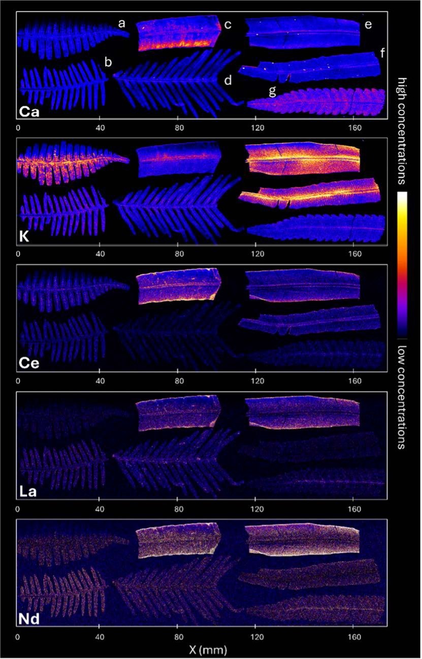
Figure 1. Shows MicroXRF elemental maps from the Atlas X of selected fern specimens, comparing calcium, potassium, cerium, lanthanum, and neodymium. Warmer colors indicate higher concentrations. Results reveal differences among specimens and that rare earth elements follow tissue-related patterns rather than hotspots. The specimens a–g are seven fern samples from the Queensland Herbarium chosen for further analysis.
The study confirmed substantial rare earth element accumulation in several selected species. Reported total concentrations included approximately 978 µg/g in Diploblechnum neglectum, 1,130 µg/g in Sticherus flabellatus var. flabellatus, and 1,290 µg/g in Sticheropsis milnei. Researchers also validated previously reported accumulation in Dicranopteris linearis and Blechnopsis orientalis, with the latter reaching approximately 3,850 µg/g total rare earth elements.
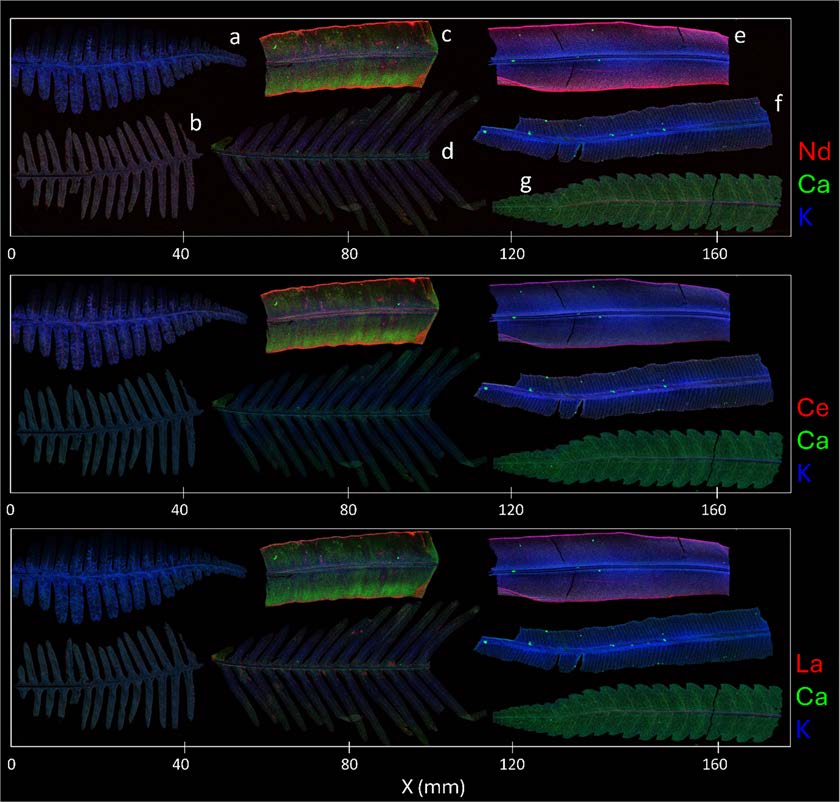
Figure 2. Presents composite microXRF maps from the Atlas X illustrating the distribution of rare earth elements relative to fern structure. Each row depicts a specific rare earth element in red—neodymium at the top, cerium in the middle, and lanthanum at the bottom—while calcium appears in green and potassium in blue. These overlays facilitate comparison between rare earth element locations, major-element patterns, and identifiable features of the plant tissue.
Why the Discovery Matters
The research demonstrates how natural-history collections can contribute to several emerging areas of science.
Herbarium specimens may help researchers identify plants that can serve as biogeochemical indicators, pointing toward soils or geological formations enriched in rare earth elements. Hyperaccumulator species may also have potential applications in phytoremediation, where plants remove elements from affected soils, and phytomining, where metal-rich biomass could eventually become a recoverable resource. The authors conclude that herbarium XRF screening may support both mineral bioprospecting and future phytomining research.
The study also illustrates the value of a tiered analytical workflow:
Rapid XRF screening identifies unusual specimens. Laboratory microXRF reveals elemental distribution. Complementary analytical methods confirm the result.
By connecting botanical collections, plant science, and critical-mineral research, this work shows how spatially resolved elemental analysis can uncover new information from specimens collected generations ago.
From preserved fern fronds to potential new pathways for rare earth element discovery—that is Atlas in Action.
Researchers and Institutions
The research was conducted by Amelia Corzo-Remigio, Imam Purwadi, Nathan Fox, Peter D. Bostock, Carlos Martel, Antony van der Ent, and Peter D. Erskine. Participating institutions included The University of Queensland Sustainable Minerals Institute, The University of Sriwijaya, Queensland Herbarium & Biodiversity Science, Royal Botanic Gardens Kew, and Wageningen University & Research.