Estimated read time: ~5–6 minutes.
Introduction: Expanding the Toolkit for Microplastic Research
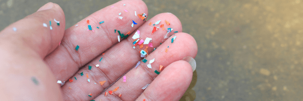
Microplastics, defined as particles smaller than 5 mm, have rapidly emerged as one of the most pervasive environmental contaminants across terrestrial, aquatic, and atmospheric systems. While techniques like Raman spectroscopy and FTIR dominate polymer identification workflows, a critical gap remains: elemental characterization of microplastics and their associated contaminants.
Researchers at the University of Memphis, through the Center for Applied Earth Science and Engineering Research (CAESER), are addressing this gap by evaluating micro-X-ray fluorescence (μXRF) as a complementary analytical tool.
Their recent work, presented at the Geological Society of America (GSA) Triple Joint Conference, explores how IXRF’s ATLAS X μXRF spectrometer can enhance microplastic detection and characterization workflows.
Research Overview: Why MicroXRF?
Traditional microplastic workflows rely on:
- Density separation
- Filtration onto glass fiber filters
- Optical microscopy
- Spectroscopic identification (FTIR, Raman)
While effective for polymer identification, these approaches do not routinely capture elemental signatures, which can provide insight into:
- Pigments and additives
- Sorbed metals and contaminants
- Environmental interactions
The University of Memphis team set out to evaluate whether μXRF could fill this gap as a non-destructive, spatially resolved elemental mapping technique.
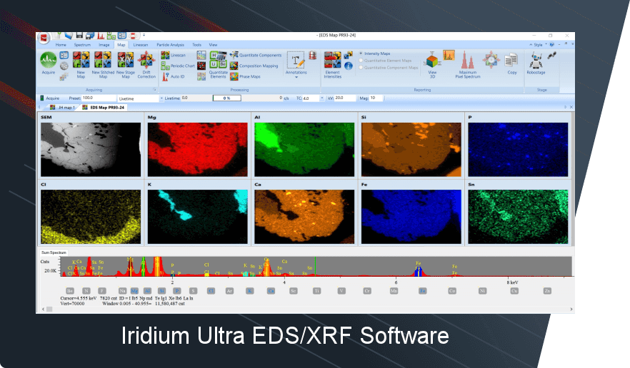
Figure 1. Scientific poster from the University of Memphis evaluating micro–X-ray fluorescence (μXRF) as a complementary method for microplastic identification. In-house microplastics and organic controls were deposited on glass fiber and stainless-steel filters and analyzed using an IXRF ATLAS X μXRF system (5–10 μm resolution). Elemental mapping reveals localized enrichments in Ti, Ca, Fe, and Cl in microplastics and highlights the absence of Si relative to the filter, enabling particle discrimination. Results demonstrate μXRF as a non-destructive tool for spatial characterization and screening of microplastics, with limitations for particles <10 μm and polymer identification.
Methodology: Real-World Microplastic Simulation
The research team designed controlled laboratory experiments using:
- In-house generated microplastics: fibers, films, foams, and fragments
- Control organic materials: cotton, wood, hair
- Substrates: 1.6 μm glass fiber filters and stainless-steel filters
Using the IXRF ATLAS X μXRF system, they performed:
- Elemental mapping at 5–10 μm spatial resolution
- 50 ms dwell time per pixel
- Comparative analysis between plastics, organics, and filter backgrounds
This approach allowed the team to evaluate both detection capability and material discrimination.
Key Findings: Elemental Signatures Unlock New Insights
1. Microplastics Exhibit Distinct Elemental Enrichment
Many microplastic particles showed elevated levels of:
- Titanium (Ti)
- Calcium (Ca)
- Iron (Fe)
- Chlorine (Cl)
These elements are commonly associated with:
- Pigments (e.g., TiO₂)
- Fillers
- Additives used in polymer manufacturing
2. Silicon Contrast Enables Particle Discrimination
One of the most powerful findings was the ability to distinguish microplastics from the silica-rich glass fiber filter background:
- Plastics: absence of Si signal
- Filter: strong Si signal
This contrast significantly improved visualization and particle segmentation, even without direct polymer identification.
3. Ratio Mapping Enhances Detection
Elemental overlays and ratio maps further improved:
- Particle visibility
- Spatial resolution of features
- Differentiation from organic materials
4. Detection on Alternative Substrates
A preliminary test using a stainless-steel filter demonstrated:
- Successful detection of microplastic fibers via localized Ti signals
- Suppression of background Fe interference
This highlights the importance of substrate selection in analytical performance.
Limitations: Where MicroXRF Fits in the Workflow
The study clearly positions μXRF as a complementary, not standalone, technique.
Key limitations include:
- Reduced sensitivity for particles <10 μm
- Inability to directly identify polymer type
- Potential for elemental overlap (e.g., Ca, Cl in both plastics and organics)
- Longer scan times for high-resolution mapping
Despite these constraints, the results demonstrate strong potential for:
- Pre-screening samples
- Supporting particle counting workflows
- Characterizing additives and contaminants
Strategic Insight: Why This Matters for Environmental Research
Microplastic research is shifting from “what is it?” (polymer ID) to “what does it carry and how does it behave?”
This is where μXRF becomes strategically valuable:
- Enables elemental fingerprinting of additives
- Supports contaminant transport studies
- Adds spatial context to particle analysis
- Enhances multi-modal workflows (XRF + FTIR/Raman)
The University of Memphis work underscores a broader trend:
Integrated analytical workflows are the future of environmental microplastic research.
See What MicroXRF Can Reveal in Your Microplastic Samples
If your research involves:
- Microplastics and environmental contamination
- Additive and pigment characterization
- Particle mapping and spatial analysis
IXRF’s ATLAS series microXRF systems can help you expand beyond traditional spectroscopy.
? Request a sample analysis or live demonstration
? Speak with an applications scientist about your workflow
FAQ Section
What is microXRF used for in microplastic research?
- MicroXRF is used to map elemental composition, helping identify additives, pigments, and contaminants associated with microplastic particles.
Can microXRF identify polymer types?
- No. MicroXRF cannot determine polymer composition directly; it is best used alongside FTIR or Raman spectroscopy.
What are the advantages of microXRF for microplastics?
- It provides non-destructive, spatially resolved elemental mapping and can highlight additives and environmental interactions.
What size microplastics can microXRF detect?
- Detection is most effective for particles larger than ~10 μm, depending on instrument configuration and scan parameters.
Acknowledgments
Special thanks to Prof. Gary E. Stinchcomb and the authors, Rodrigo Villalpando-Vizcaino, Cristina Leschhorn, and Daniel Larsen, for sharing their research and advancing the field of microplastic analysis. Their work highlights the growing importance of integrating elemental analysis into environmental workflows and reinforces the role of microXRF as a powerful complementary tool.