Synchrotron-based X-ray fluorescence microscopy (XFM) has proven to be an invaluable resource in medical research, offering unparalleled levels of contrast and precision while studying complex microstructures. However, with limited access due to high costs and lengthy wait times for technicians and scientists, it can take years before research is performed and results are obtained. This article examines the reliability of laboratory-based micro X-ray fluorescence (microXRF) systems as a cost-effective alternative, referencing their current application in medical research laboratories worldwide. Compared to traditional Synchrotron methods, our study highlights how such offerings dramatically decrease wait times while providing comparable accuracy – allowing researchers from academia and the medical industry greater XRF access and faster results.
Laboratory microXRF systems offer a practical and more accessible alternative to XFM beamlines for various research applications. Although they typically provide lower spatial resolution and sensitivity compared to synchrotron-based techniques, microXRF systems can still be highly valuable for various studies. Here are some ways in which a laboratory microXRF system with a 5-micron X-ray beam size could assist:
Preliminary and Feasibility Studies
- Screening Samples: Quickly assess and screen a large number of samples to identify promising candidates for more detailed synchrotron-based analysis.
- Optimizing Experimental Conditions: Develop and optimize experimental protocols and conditions before using valuable beamline time.
Accessibility and Availability
- No Beamtime Constraints: Unlike synchrotrons requiring proposal submission and allocated beamtime, laboratory systems are much more accessible and can be used on demand.
- In-House Experiments: Conduct experiments in-house without the need for travel or transport of delicate samples.
Spatial Resolution
- Adequate Resolution for Many Applications: A 5-micron beam size is sufficient for various applications, mainly when ultra-high spatial resolution is not critical.
Complementary Data Collection
- Broad Elemental Range: Laboratory systems can detect a wide range of elements, providing complementary data to synchrotron XFM.
- Multiple Modalities: Some microXRF systems offer additional imaging or analytical capabilities, such as X-ray absorption contrast imaging or X-ray diffraction.
Training and Education
- Hands-On Experience: Provide training and hands-on experience for students and researchers in XRF techniques and data analysis.
- Method Development: Develop and refine methodologies for XRF analysis and interpretation.
Time-Efficient Studies
- Rapid Data Acquisition: Laboratory systems can offer rapid data acquisition times for certain samples and analyses.
- Immediate Feedback: Obtain immediate results and feedback, allowing for iterative experimentation.
Cost-Effectiveness
- Reduced Expenses: Avoid the significant costs associated with travel, shipping of samples, and beamline usage fees.
- Resource Optimization: Optimize the use of synchrotron beamtime by conducting preliminary and complementary studies in-house.
Flexibility in Sample Preparation
- Diverse Sample Types: Laboratory systems may accommodate a broader range of sample types and sizes.
- Iterative Sample Preparation: Quickly adjust sample preparation techniques based on immediate results.
Case Study
In a recent study, we worked alongside Tatjana Paunesku and Jeffery Murley from Northwestern University to conduct a comparative analysis of data gathered using Synchrotron-based X-ray Fluorescence Microscopy (XFM) and laboratory-based microXRF, highlighting the high spatial resolution offered by the latter.
The original study utilized XFM to analyze formalin-fixed paraffin-embedded (FFPE) lung and lymph node tissues from a beagle exposed to 90Y-aluminosilicate microparticles in 1968. The spatial distribution of elements, including Si (from microparticles), P, S, Fe, and Zn in the tissues, was mapped using XFM. Aluminosilicate microparticles were detected in both tissues, with a higher concentration in the lymph node. The study confirmed the co-localization of Si and Fe and observed that in some high-resolution images, Fe signals were adjacent to but not fully overlapping with Si signals. The study also detected Zr, the decay product of 90Y, in the lymph node, suggesting the stability of microparticles inside cells during transport. The original study highlighted the potential of XFM in investigating archival samples for markers of internal radiation exposure.
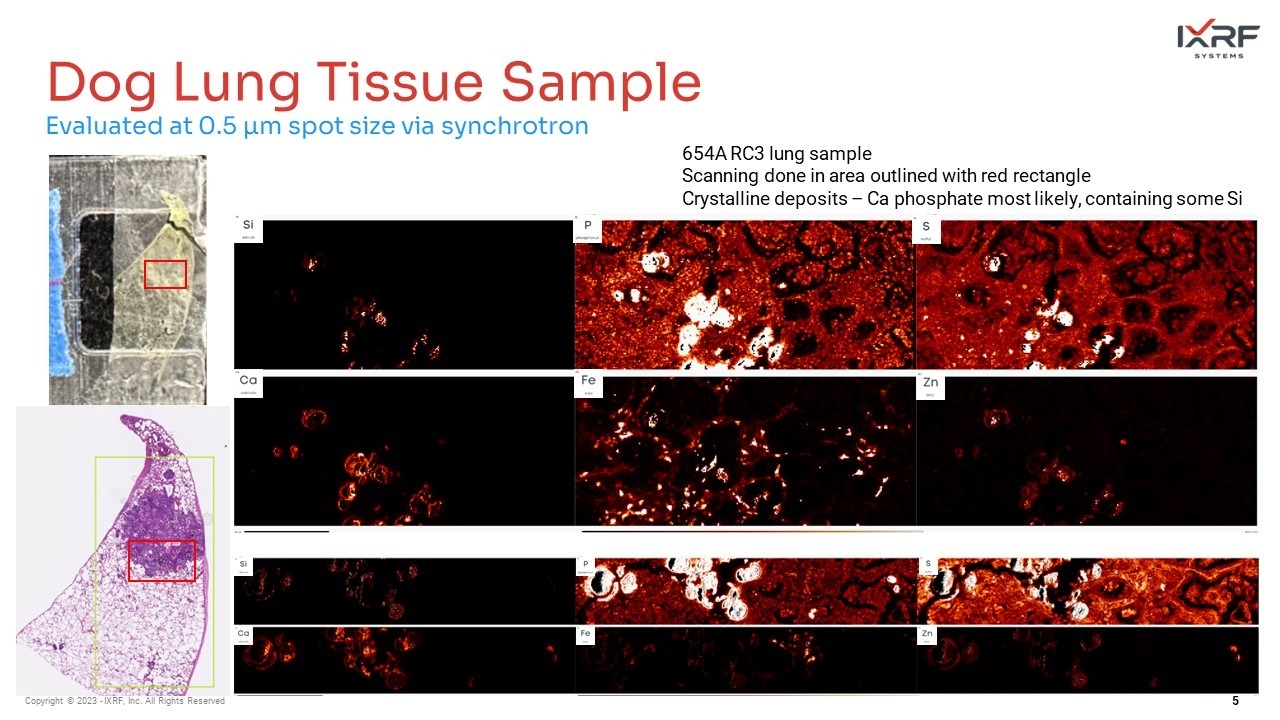
Figure 1: XFM elemental maps of dog lung tissue sample showing the distribution of Si, P, S, Ca Fe, and Zn. Maps were collected using an X-ray beam spot size of 0.5 μm and 10.5 keV beam energy.
Our study analyzed a sample from the original study using the Atlas X microXRF, equipped with a polycapillary X-ray source that provides a 5 µm spot size. Our findings indicate that the laboratory-based microXRF is a valuable tool for generating elemental maps, which helped us identify regions enriched with Si and elevated levels of P, S, Ca, and Fe for further investigation. As a result, this technology has proven effective in performing a significant portion of the initial low-resolution mapping and analyses required in our study, paving the way for more targeted and nuanced investigations.


Figure 2: (1) Laboratory-based MicroXRF elemental maps show the distribution of Si, P, S, Ca, and Fe. (2) Merged elemental maps show the overlayed distribution of elements without colocalization. (3) Sub-map region of interest collected, providing a close-up evaluation. Maps were collected using a 5 µm spot size.
Conclusion
Integrating laboratory microXRF systems, particularly those with a 5-micron beam size, plays a crucial role in complementing the unique capabilities of synchrotron XFM beamlines. While it may not be a direct replacement, it significantly broadens the horizons of research applications across various fields. Its accessibility ensures that research can be conducted without requiring extensive resources or specialized facilities. At the same time, its flexibility allows for a diverse array of applications, catering to the specific needs of different research projects. Additionally, the cost-effectiveness of laboratory microXRF systems makes advanced analytical techniques more attainable, breaking down financial barriers and democratizing access to high-quality data.
In the context of biomedical research, the high spatial resolution provided by laboratory-based microXRF systems opens new avenues for exploration and analysis. By enabling the detailed mapping of elemental distributions within biological tissues, researchers are equipped with the tools to uncover intricate cellular processes and interactions at a microscale. This enhanced understanding can lead to more precise diagnostics, targeted therapies, and personalized medicine, ultimately contributing to advancements in healthcare and patient outcomes. In this way, laboratory-based microXRF with high spatial resolution acts as a catalyst for innovation and progress in biomedical research, complementing and enhancing the capabilities provided by synchrotron XFM beamlines.
Discover the practical applications and benefits of the Atlas series microXRF for your research. We invite you to contact us for more detailed information and to discuss how this technology can be tailored to meet the specific needs of your projects. Whether you are engaged in biomedical research or other scientific fields, the Atlas series microXRF offers high spatial resolution and precision in elemental analysis. Don’t hesitate to reach out and learn more about how this tool can contribute to the efficiency and accuracy of your work. We are here to answer your questions and assist you in making an informed decision on integrating this technology into your research toolkit. Contact us today to enhance your research capabilities with the Atlas series microXRF .
IXRF Systems is a leading provider of X-ray fluorescence instrumentation. With our advanced analytical solutions, we are committed to supporting research, quality control, and educational endeavors across various industries. Click here to contact us today to get a deeper understanding of how our technology can be a valuable addition to your analytical toolkit.