MicroXRF for Biological and Environmental Elemental Mapping
Atlas Apex provides non-destructive microXRF analysis of soil, plant tissue, biological specimens, filters, particulates, and other heterogeneous environmental samples.
Unlike bulk methods that report only an average concentration, microXRF shows where elements are located across the sample. Researchers can map nutrients, trace elements, heavy metals, and contaminants while preserving valuable spatial information.
With the appropriate detector configuration and measurement environment, Atlas Apex supports elemental analysis from carbon through uranium.
Why Spatial Elemental Analysis Matters
Where microXRF fits: Atlas Apex is best suited for non-destructive elemental mapping, hotspot detection, and region-of-interest discovery. It complements bulk methods such as ICP-MS, higher-resolution techniques such as SEM-EDS, and molecular methods such as FTIR and Raman.
Biological and environmental samples are rarely uniform.
Bulk analysis can determine how much of an element is present, but it cannot show where that element occurs.
A digested leaf sample may confirm total zinc concentration, for example, but it cannot show whether the zinc is concentrated in vascular tissue, outer tissue, or a localized deposit. Similarly, a homogenized soil sample may indicate elevated lead without revealing whether it is associated with the surface, a particular layer, or individual particles.
MicroXRF preserves this spatial context by mapping the original sample rather than averaging it. It can help researchers distinguish:
- Uniform concentrations from localized hotspots
- Surface deposits from internal accumulation
- Tissue-specific uptake from whole-sample averages
- Contamination layers within soil or sediment
- Individual particles from broadly distributed material
Life Science and Environmental MicroXRF Maps
Plant Tissue and Botanical Samples
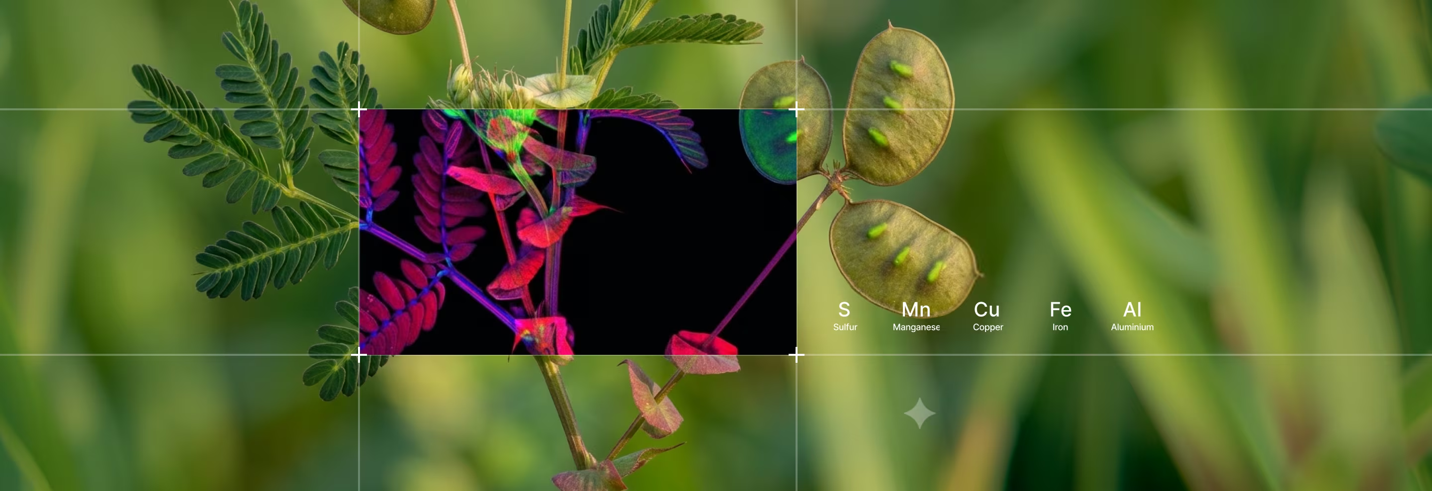
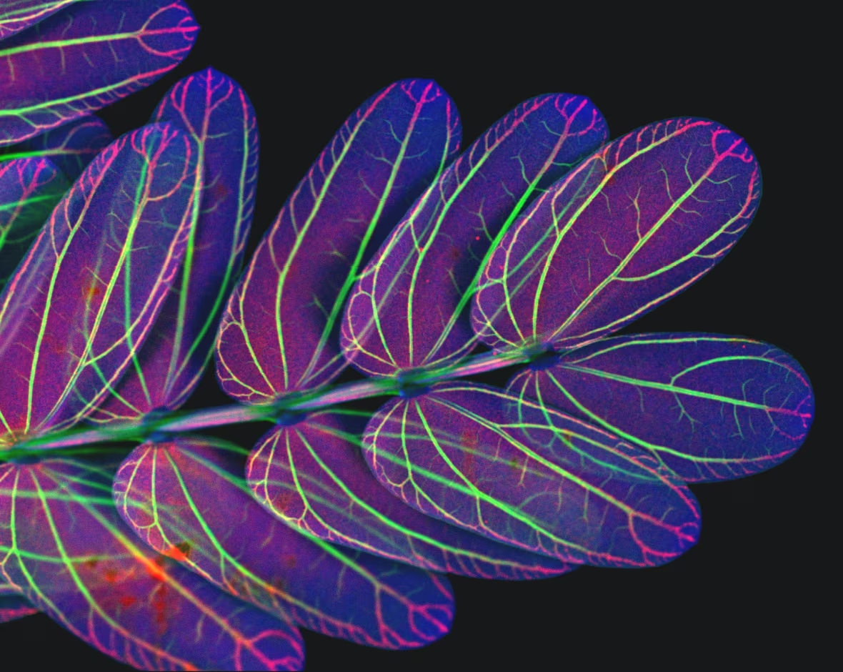
MicroXRF maps elemental distribution across leaves, stems, roots, seeds, and botanical cross-sections without reducing the specimen to a single bulk measurement. Researchers can see where nutrients, trace elements, and absorbed metals accumulate and how their distributions vary across plant structures.
MicroXRF can help researchers study:
- Nutrient transport and storage
- Deficiency and toxicity
- Uptake from soil or irrigation water
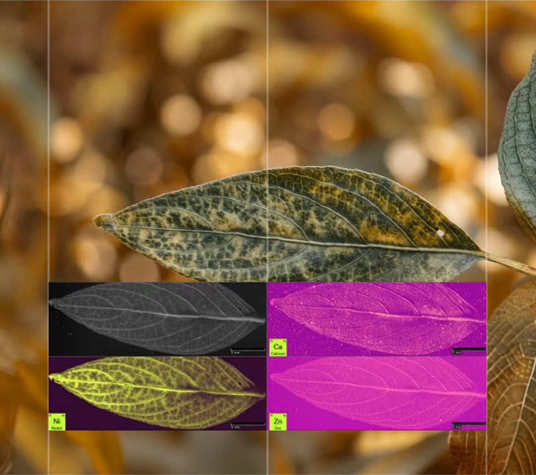
- Tissue-specific metal accumulation
- Differences between exposed and control plants

Revealing Tissue-Specific Metal Accumulation
MicroXRF is especially useful for studying hyperaccumulator species and plants exposed to contaminated soil. It can show where absorbed metals are concentrated within the tissue and whether they follow veins, margins, growth structures, or localized deposits.
Biological Tissue and Specimens
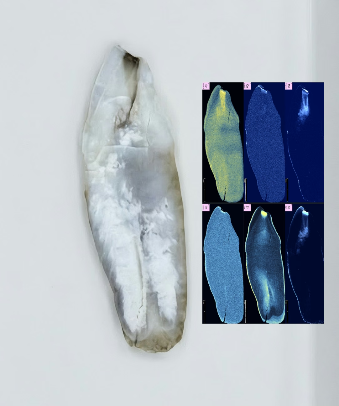
Elemental distribution can provide insight into biological development, environmental exposure, diet, and mineralized structures.
Suitable samples may include:
- Tissue sections
- Bone and teeth
- Shells and otoliths
- Invertebrate specimens
- Preserved biological materials
- Mineralized tissue
Elemental maps can reveal growth banding, localized metal accumulation, and chemical differences across tissue layers.
Toxicology and Exposure Studies
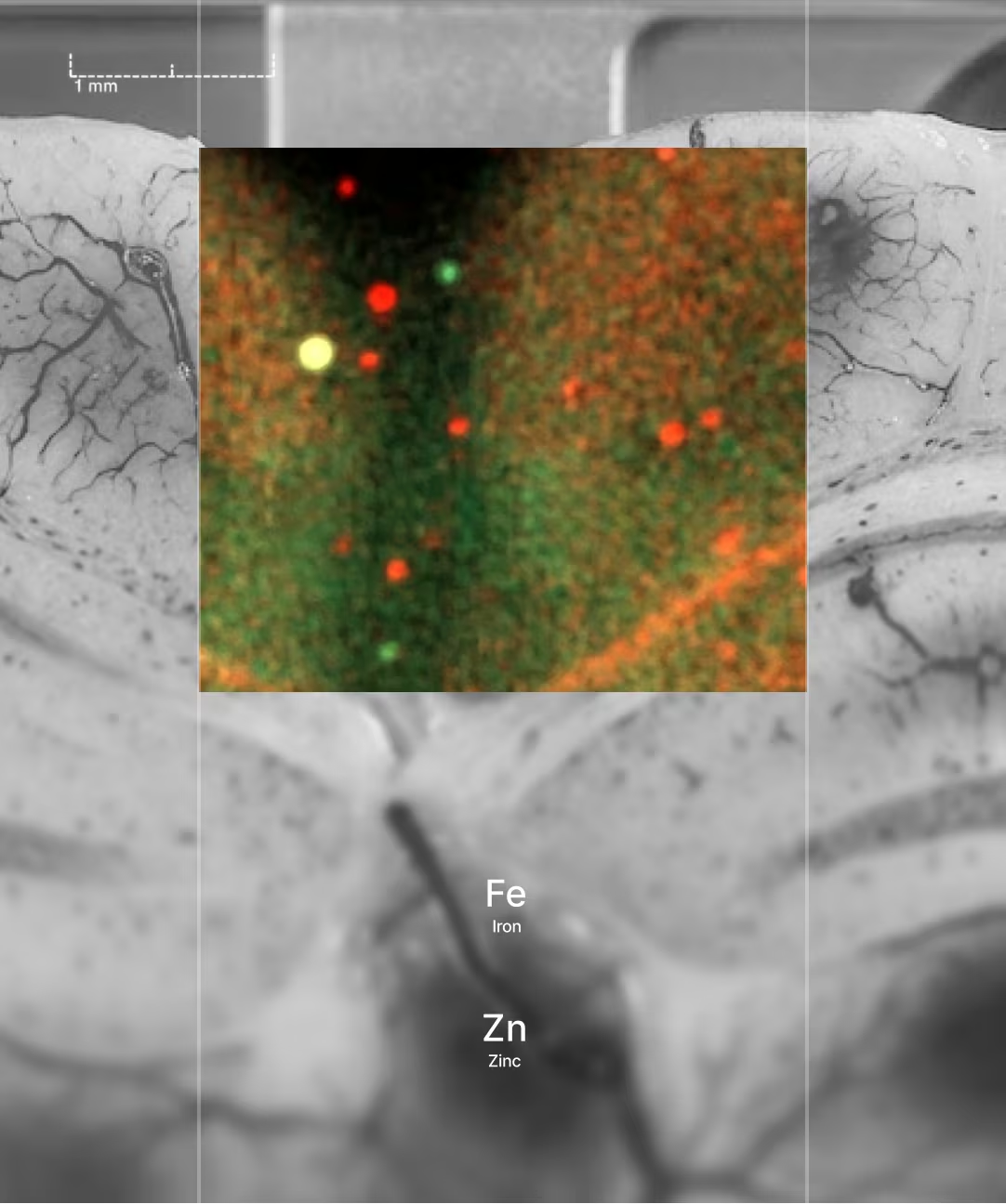
Toxicology research often requires more than confirming that an element entered an organism. Researchers also need to know where it accumulated.
MicroXRF can help investigate:
- Uptake pathways
- Tissue-specific accumulation
- Exposure gradients
- Element transport
- Co-localization of multiple elements
- Differences between exposed and control specimens
Environmental Applications
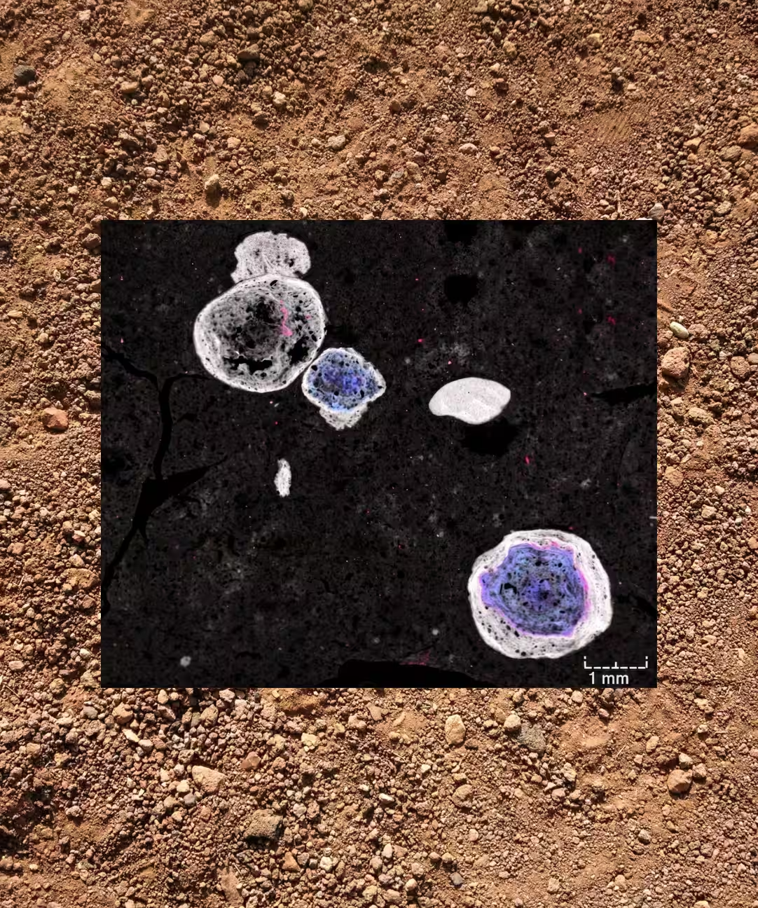
Soil and Sediment Analysis
Contaminants in soil and sediment are rarely distributed uniformly. They may be concentrated near the surface, associated with a particular layer, or attached to specific grains and mineral phases.
MicroXRF can map:
- Lead, arsenic, chromium, and nickel
- Copper, zinc, cadmium, and mercury
- Industrial or mining-related metals
- Contamination depth and element-bearing particles
- Associations between contaminants and mineral phases
Water Filters & Suspended Particulates
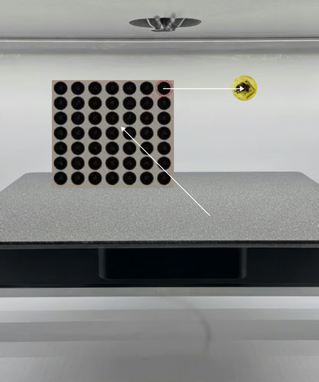
Particles collected from water can concentrate metals and other elements that may be difficult to detect in the surrounding liquid. Their composition, size, and spatial distribution can provide important clues about contamination sources, transport pathways, and environmental exposure.
MicroXRF can map:
- Metals captured on membrane and cartridge filters
- Iron, copper, zinc, lead, arsenic, and other contaminants
- Individual mineral or industrial particles
- Elemental associations within suspended material
- Localized hotspots that may be diluted in bulk analysis
Air Particulates
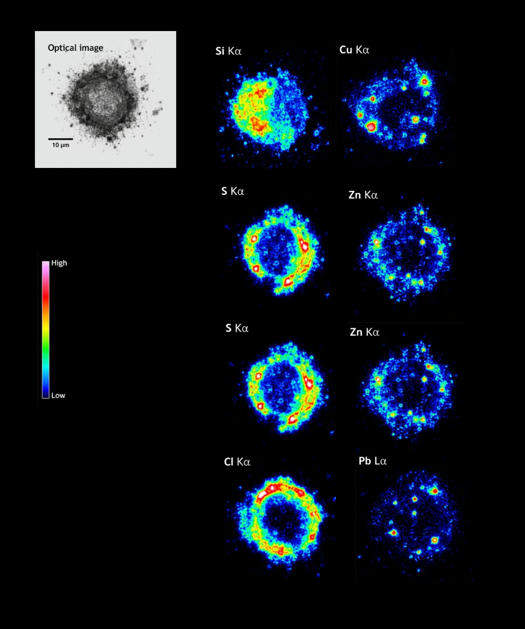
Airborne particles can originate from natural dust, combustion, industrial processes, mining, transportation, and other environmental sources. Bulk measurements may determine total elemental loading, but they cannot show whether a signal comes from many similar particles or a small number of highly enriched contaminants.
MicroXRF can map:
- Metal-bearing airborne particulates
- Combustion- and industrial-related elements
- Mineral dust and soil-derived particles
- Deposited particles collected on air filters
- Elemental hotspots and particle-to-particle variation
Microplastics & Elemental Tracers
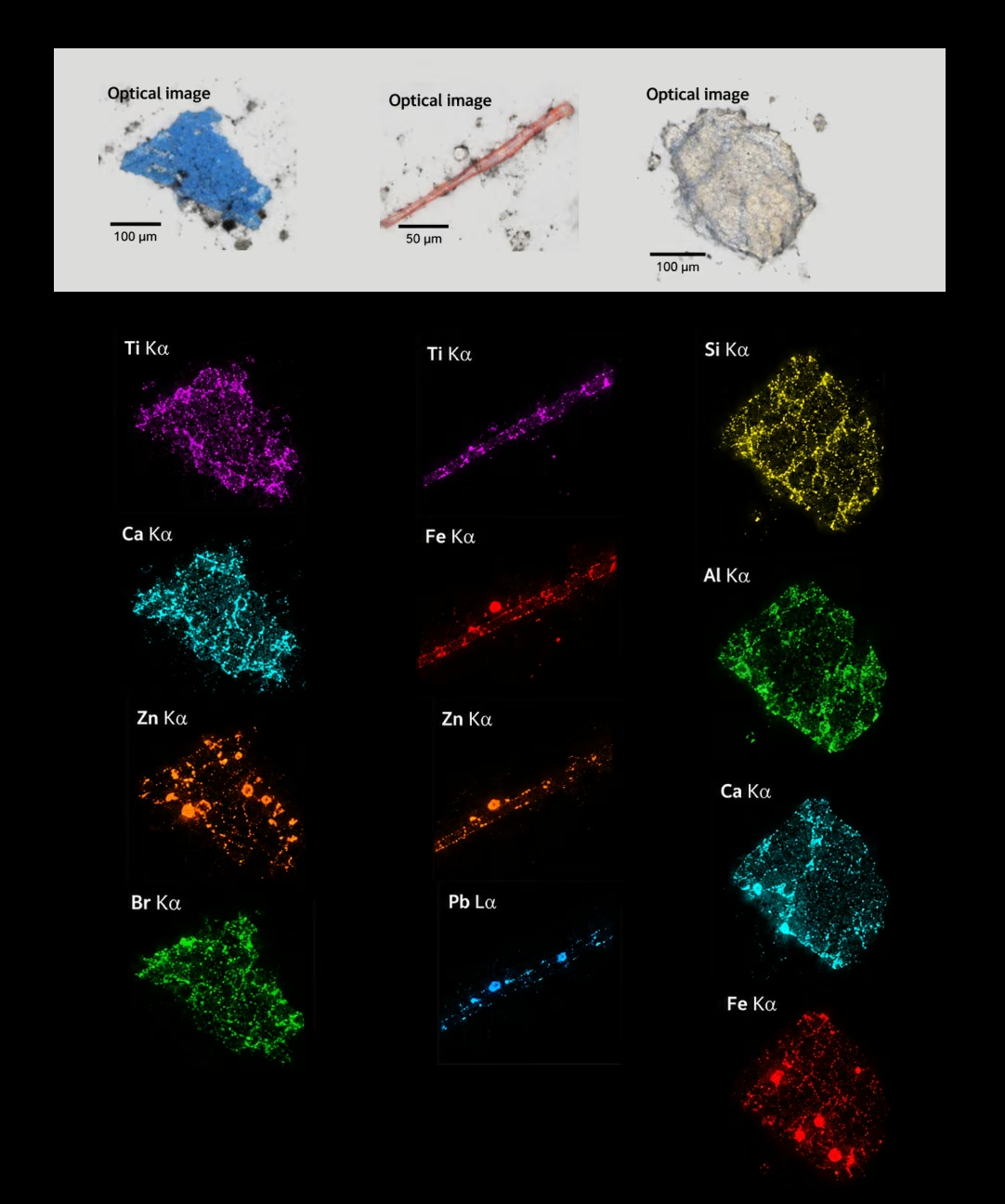
Microplastics can contain inorganic fillers, pigments, additives, coatings, and contaminants that provide elemental signatures useful for characterization. Environmental particles may also acquire metals or mineral material during transport through water, soil, sediment, or biological systems.
MicroXRF can map:
- Inorganic pigments and fillers within plastic particles
- Metal-containing additives and coatings
- Elements associated with weathered particle surfaces
- Adsorbed environmental contaminants
- Elemental tracers used to distinguish particle populations
How Atlas Apex MicroXRF Analysis Works
Atlas Apex combines focused X-ray excitation, automated sample positioning, large-area mapping, and hyperspectral data collection to examine biological and environmental samples from the full specimen down to localized elemental features.
Prepare and Position the Sample
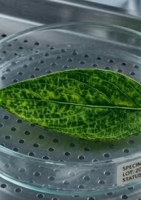
Samples may be analyzed as intact specimens, tissue sections, soil or sediment cores, mounted particles, powders, or material collected on filter media.
Conductive coating, chemical digestion, and ashing are not required for many applications. Integrated overview and spot-view cameras help users locate the sample, document its condition, and select points, lines, maps, or regions of interest.
Examine Features with Focused-Beam Analysis
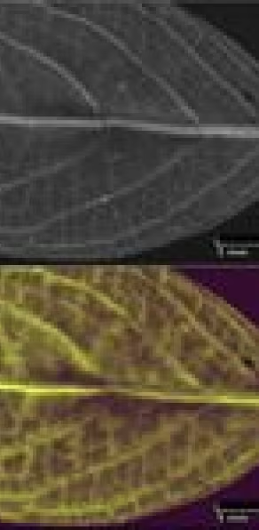
Available Atlas Apex configurations provide a nominal X-ray spot size as small as approximately 5 µm, helping researchers investigate:
- Tissue layers and growth structures
- Small areas of elemental accumulation
- Individual particles and soil grains
- Vascular structures in plant samples
- Localized deposits and contamination
Map Large Samples and Complete Regions of Interest
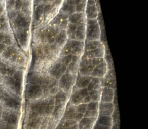
Atlas Apex combines focused analysis with an automated mapping area of up to approximately 220 × 200 mm. Researchers can map complete tissue sections, filters, plant specimens, and soil cores rather than relying on isolated spot measurements — connecting small elemental features with their position across the full specimen.

Collect a Complete Spectrum at Every Pixel
Researchers can generate maps for additional elements, compare distributions and overlap, investigate unexpected signals, and reprocess data without rescanning the sample. Iridium Ultra integrates acquisition, spectral processing, mapping, and quantitative analysis.
Support Flexible and In-Situ Experiments
Samples can be analyzed in air for many routine applications; vacuum or helium conditions improve sensitivity to lighter elements when the sample is compatible. A rear chamber access port supports compatible accessories, experimental fixtures, and user-developed sample environments for in-situ or controlled experiments.
MicroXRF Compared With Other Analytical Methods
vs. ICP-MS or ICP-OES
ICP-MS and ICP-OES provide precise bulk concentrations and very low detection limits, but they normally require sample digestion. MicroXRF preserves the original sample and adds spatially resolved elemental information — complementary when researchers need both bulk concentration and elemental distribution.
vs. Atomic Absorption Spectroscopy
Atomic absorption spectroscopy generally provides an averaged concentration for selected elements after sample preparation or digestion. MicroXRF adds the ability to locate elemental hotspots, gradients, layers, and individual particles.
vs. SEM-EDS
SEM-EDS provides higher spatial resolution and detailed electron imaging, but it generally examines a smaller, prepared sample. MicroXRF accommodates larger samples, requires less preparation, and maps wider areas without a conductive coating — and can identify regions of interest for subsequent SEM-EDS examination.
with FTIR or Raman Spectroscopy
FTIR and Raman identify molecular structures and are commonly used to confirm polymer composition. MicroXRF provides complementary elemental information about pigments, fillers, flame retardants, additives, and adsorbed metals.
See What MicroXRF Can Reveal in Your Samples
Describe your sample type, elements of interest, and research objective to an IXRF applications scientist. We can help determine whether microXRF is appropriate, recommend an Atlas Apex configuration, and discuss a sample feasibility study.
Atlas Apex systems are designed and manufactured in Austin, Texas, with applications and technical support provided directly by IXRF Systems.
Frequently
Asked Questions
MicroXRF is used to map the spatial distribution of nutrients, trace elements, heavy metals, and contaminants in biological and environmental samples. Common applications include plant tissues, soils, sediments, filters, particles, and interfaces where elemental location matters as much as overall concentration.
Yes. MicroXRF can show where elements accumulate within roots, stems, leaves, seeds, and other plant tissues. This can help researchers study nutrient transport, metal uptake, hyperaccumulation, toxicity, and interactions between plants and their growing environment.
Yes, in some cases. Fresh or hydrated tissue can be analyzed when it is appropriately contained and compatible with the measurement environment. Moisture, thickness, sample stability, and geometry can affect X-ray transmission, spatial resolution, and quantitative accuracy, so sample preparation should be matched to the research objective.
Yes. MicroXRF is particularly useful for heterogeneous environmental samples because it can map elemental distributions across soil–root interfaces, sediment layers, particulate deposits, filter media, and individual particles. This helps reveal localized enrichment and chemical gradients that may be lost in bulk analysis.
MicroXRF can detect and map many environmentally important metals and metalloids, including lead, arsenic, chromium, nickel, copper, zinc, cadmium, and mercury. Detectability depends on concentration, sample matrix, particle size, preparation, and measurement conditions.
Yes, but microXRF does not directly identify the polymer itself. It can detect pigments, fillers, inorganic additives, flame-retardant elements, and metals adsorbed onto or associated with suspected microplastic particles. FTIR or Raman spectroscopy is typically used when molecular confirmation of the polymer is required.
Not necessarily. Many samples can be analyzed as intact sections, cores, filters, mounted particles, or prepared biological specimens without digestion. Drying, grinding, polishing, or other preparation may improve reproducibility or quantitative accuracy depending on moisture, geometry, and the analytical objective.
Yes. MicroXRF can provide quantitative elemental information when appropriate calibration or Fundamental Parameters methods are used. Accuracy depends strongly on sample thickness, moisture, matrix composition, surface condition, geometry, and homogeneity, particularly for complex biological and environmental materials.
MicroXRF is generally better suited to analyzing particulate matter collected from water, such as material captured on filters, than measuring trace elements dissolved directly in water. Dissolved elemental concentrations are more commonly measured using techniques such as ICP-MS, ICP-OES, or atomic absorption.
MicroXRF can support screening, research, source investigation, and spatial characterization, but whether it can be used for regulatory compliance depends on the required method, calibration, detection limits, quality controls, and governing standard. Confirmatory testing with an approved reference method may still be required.