
Benchtop MicroXRF for Segmented Samples and Routine Research
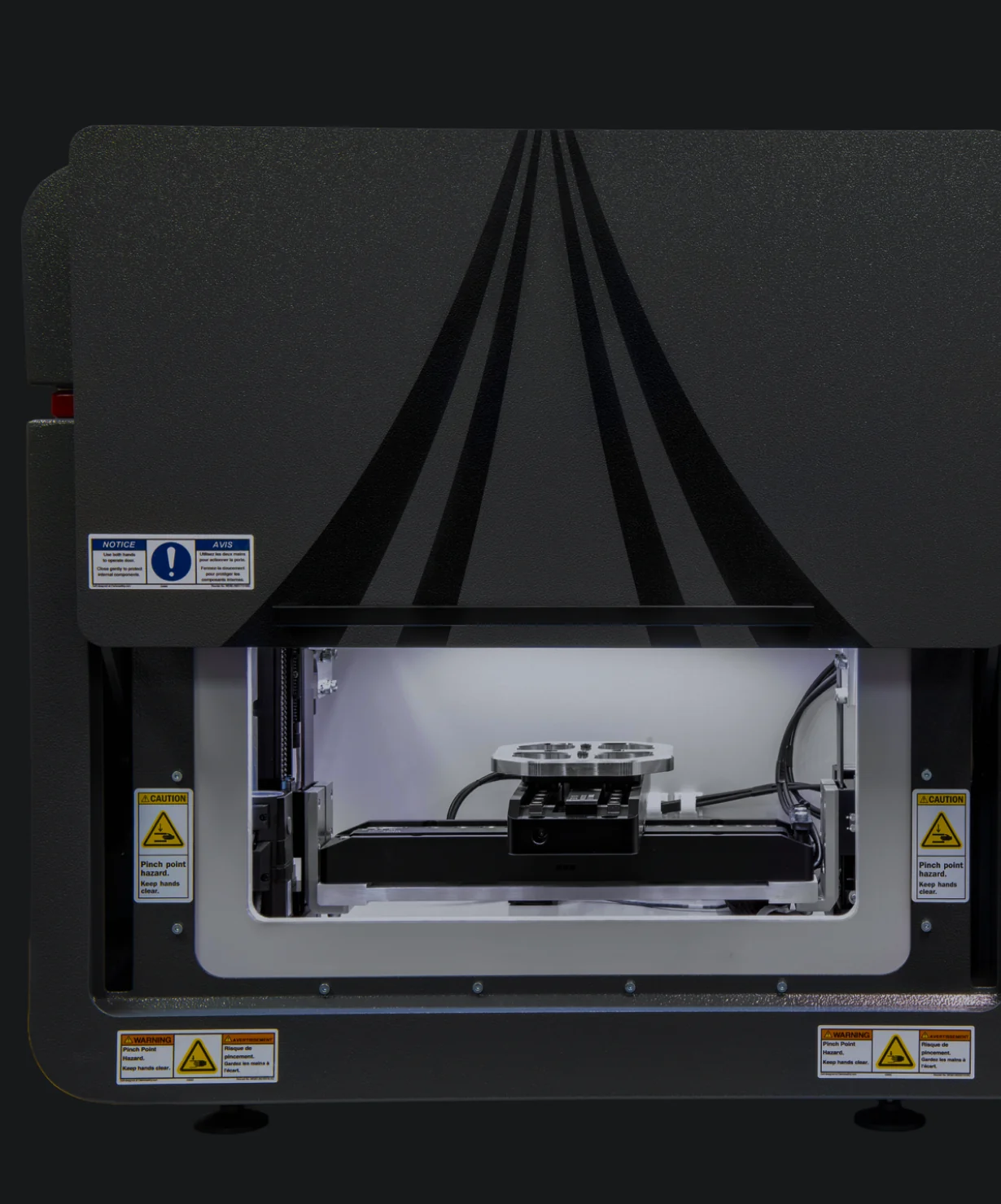
A compact benchtop microXRF spectrometer built for the same 5 µm resolution and perpendicular geometry as the rest of the Atlas Apex line, in a footprint that fits where lab space is limited.
- Smaller footprint, lower entry price
- Automated mapping across 4-inch-wide specimens
- Vacuum-ready in under one minute
- Sample chamber
- 508 × 457 × 254 mm
- Best fit
- Segmented samples,
routine research - Mapping area
- 220 × 200 mm
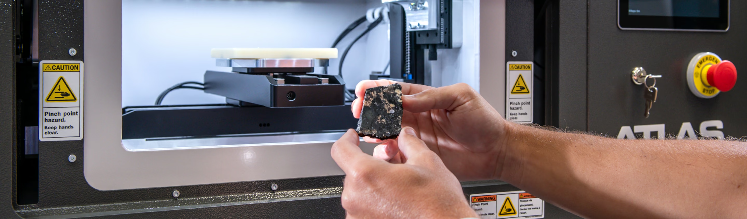
The Same Optical Engine, a Smaller Footprint
The Atlas Apex M is the compact configuration of the Atlas Apex line. It delivers the same research-grade microXRF capability as the Apex X, including spot sizes down to 5 µm and full carbon through uranium elemental coverage, in a benchtop unit that fits in labs where space is limited. For buyers who need high-spatial-resolution elemental mapping but cannot accommodate a large-area floor system, the Apex M covers that need.
Need to cover a longer core, a full wafer, or a wider variety of sample sizes instead?
What Makes Atlas Apex Different From Conventional µXRF
Not marketing claims. The specific geometry, detector, and vacuum choices that set the resolution and sensitivity ceiling, and they apply to Apex M exactly as they do to Apex X.
Perpendicular Geometry
The X-ray source is normal to the sample, producing a true circular spot down to 5 µm with no elliptical distortion. Angled-geometry competitors are limited to a 25 µm minimum spot because of beam ellipsing.
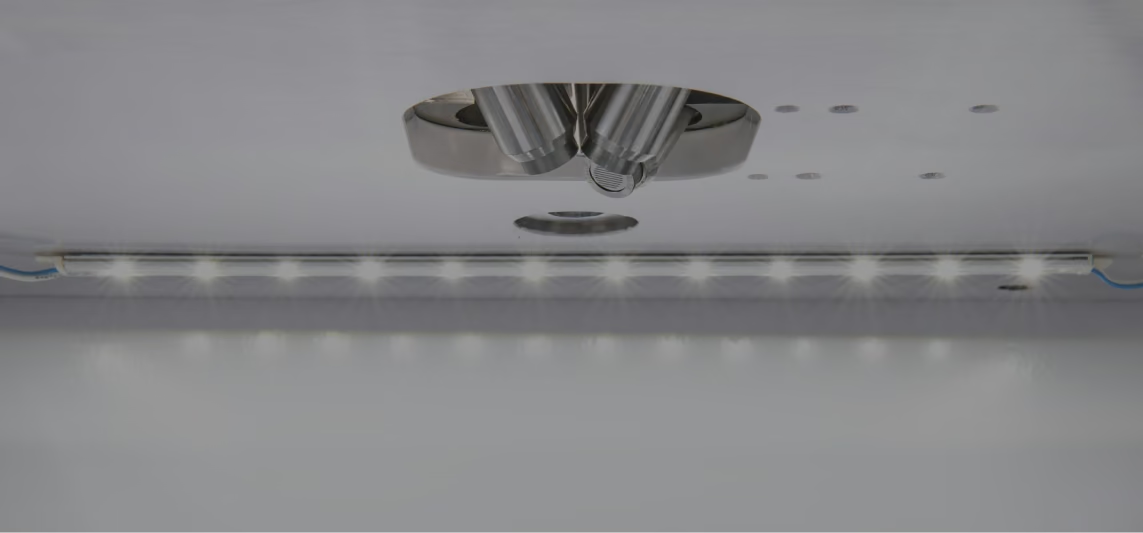
Up to Four SDD Detectors
Bundle up to four Silicon Drift Detectors for a combined active area of 260 mm², boosting throughput, sensitivity, and trace-element detection limits without compromising resolution.
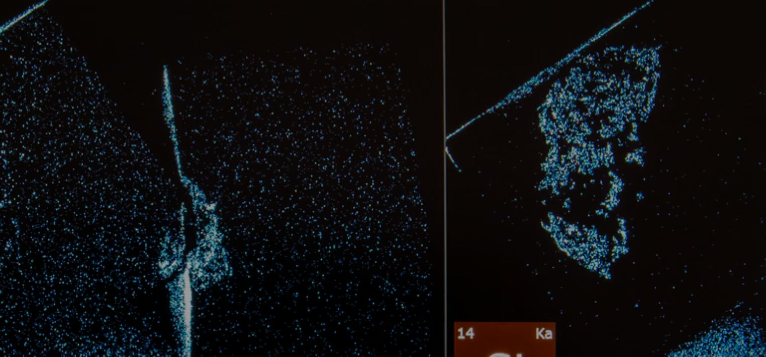
Full Spectrum, Every Pixel
Every mapped pixel stores a complete XRF spectrum. Users can generate new elemental maps, extract spectra, or re-quantify data after acquisition — without rescanning the sample.
Enhanced Vacuum Capability
Sealed chamber with adjustable vacuum pressure (<0.1 kPa) significantly boosts sensitivity for low atomic number elements, extending detection down to sodium — and carbon on the LE configuration.
“Atlas Apex microXRF delivers ~10 µm pixel resolution, revealing fine-grained mineral phases and chemical zoning that remain hidden at coarser resolutions, approaching the detail of synchrotron-based mapping.”
Atlas Apex Across Geology, Life Science, and Industry
From ore deportment to plant metal homeostasis to PCB failure analysis — real Atlas Apex output, not renders.
Ore Mineralogy and Deportment
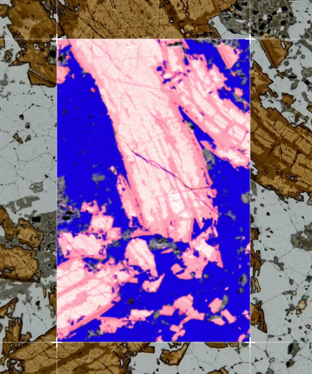
A sphalerite-bearing ore sample from the Philipsburg mining district shows a quartz vein in dolomitic carbonate host rock, with secondary Fe and Cu minerals from weathering. At pixel sizes under 20 microns, the finely intergrown nature of secondary minerals — and the polymetallic association of Cu, Fe, As, and Zn — becomes visible in ways bulk assay alone cannot show.
In gold deportment work specifically, Atlas Apex maps how gold relates to pyrite, arsenopyrite, arsenic, and sulfur — helping metallurgists understand whether gold is likely free, attached to other minerals, or locked within sulfide phases.
Phytomining and Plant Metal Homeostasis
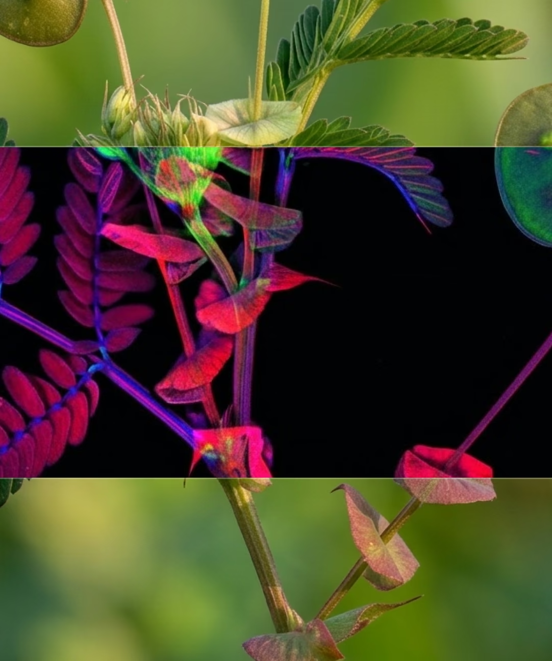
Atlas enabled high-resolution elemental mapping of selenium across whole plant structures of Neptunia amplexicaulis, revealing tissue-specific hyperaccumulation patterns that support hypotheses on phloem-mediated selenium redistribution and taproot storage.
The same technique applies across botany more broadly — from heavy-metal to REE hyperaccumulation studies, entirely non-destructive, with no coating or sample prep required.
Microelectronics: Assembly to Defect
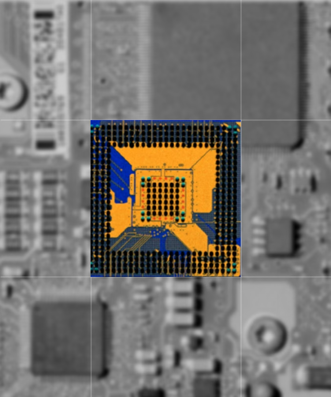
Atlas Apex connects broad-area elemental screening with micron-scale investigation on one platform — mapping material composition across boards, components, traces, and complex assemblies, then targeting anomalous features with high-resolution sub-maps.
5 µm mapping resolves fine interconnects; K/Ca overlays localize packaging residues and particles; Ag/Au overlays spatially resolve wire-bond and frame connections — all without sectioning the device.
Battery Electrodes and Energy Storage
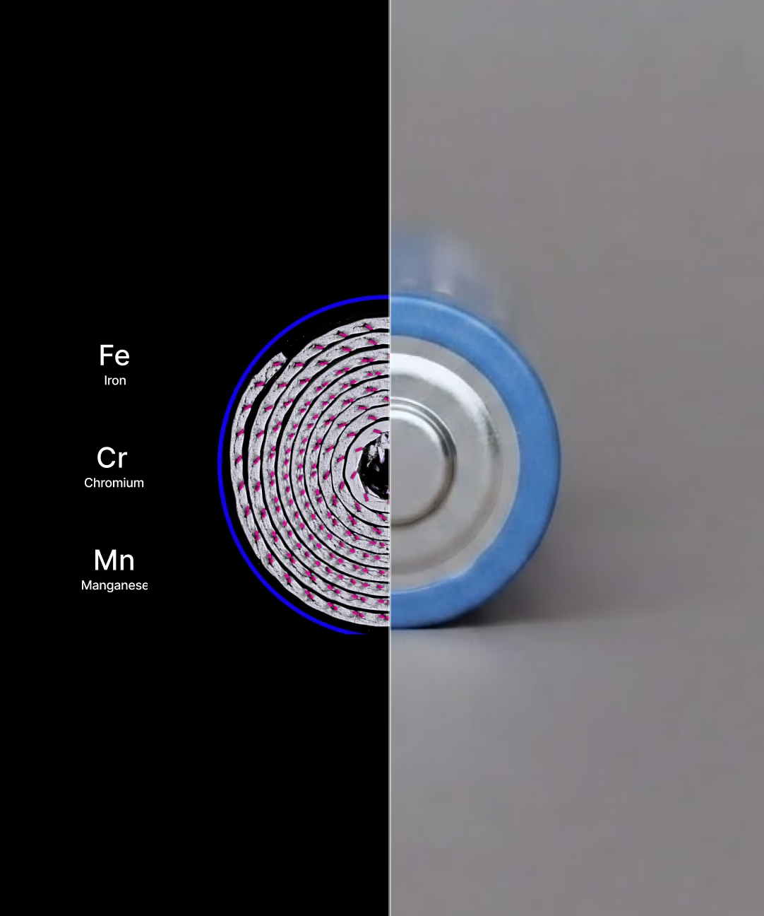
MicroXRF maps can reveal variations in electrode coating, current-collector defects, contamination, and transition-metal crossover between cell components. Depending on chemistry, elements of interest may include nickel, cobalt, manganese, iron, aluminum, phosphorus, sulfur, and copper.
Iridium Ultra™ — Hyperspectral XRF Mapping
Atlas Apex is powered by Iridium Ultra, IXRF's in-house microXRF software platform for Windows® 11. It integrates acquisition, spectral processing, quantitative analysis, hyperspectral mapping, phase classification, image analysis, and reporting into a single workflow — with every pixel in a map storing a full XRF spectrum.
Quantitative Analysis
- Fundamental Parameters analysis for solids, liquids, powders, particles
- Thin-film FP for up to 8 layers plus infinite substrate
- Automatic peak ID, overlap correction, deconvolution
- Quantitative Match and material classification database
Elemental & Phase Mapping
- Hyperspectral mapping up to 9,998 × 9,998 pixels
- Simultaneous acquisition of up to 35 elements
- Principal Component Analysis for phase mapping
- IU-GEOCHEM mineral ID against a ~4,000-composition library
Mosaic Imaging & ROI
- Optical mosaic imaging up to 100 megapixels
- Full-sample visualization beyond the native camera field of view
- Multi-point automated analysis directly from the image
- Integrated crosshair overlay for XRF/optical correlation
Linescans & Particle Analysis
- Line profile analysis across interfaces, coatings, gradients
- Tiled or stacked chart views for comparison
- Morphology and particle segmentation tools
- Automated counting and measurement by feature type
Atlas Apex M Full Specifications
Same optical engine, same software as Atlas Apex X. The four rows highlighted in red are what actually change between the two chambers; everything else is shared.
- Sample chamber size
- 508 × 457 × 254 mm
- Outside dimensions
- 890 × 560 × 560 mm
- Mapping travel
- 220 (X) × 200 (Y) mm
- Stage travel
- 320 × 320 × 210 mm
- Elemental range
- Fluorine (F) through Americium (Am); Carbon (C) through Am on LE version
- Analysis atmosphere
- Air, vacuum, or He(g) purge
- Primary X-ray source
- Rh target, 50 W max, 50 kV @ 1 mA (other anodes available)
- Spot size (primary)
- 5 µm standard (10, 15, 20, 25, 40, 100 µm optional)
- X-ray filters
- Up to 7 plus open position
- Geometry
- Top-down beam, perpendicular to sample stage
- Detectors
- 1 standard, up to 4 (260 mm² total active area)
- Spectral resolution
- ≤125 eV
- Mapping scan speed
- 1–3 ms/pixel
- Stage XY speed
- Up to 300 mm/s
- Sample view
- 3 cameras for positioning and analysis
- Software
- Iridium Ultra on Windows® 11
- Vacuum readiness
- 0.1 kPa in ~120 seconds
- Quality & safety
- CE marked, RoHS, radiation < 0.5 µSv/h
Let's Evaluate Your Samples
Confirm target applications and sample types, define the required spot size and detector configuration, and run proof-of-concept measurements. We'll help determine whether Atlas Apex M or Atlas Apex X is the right fit for your lab.
Designed and built in Austin, Texas. IXRF systems are backed by applications, engineering, training, and service support from the people who make them.
Frequently
Asked Questions
The Atlas Apex M is a chamber-based microXRF system designed for versatile laboratory analysis, while the Atlas Apex X uses a more open geometry to accommodate large, heavy, irregular, or intact samples that may not fit inside a conventional chamber. Both platforms use the Atlas Apex analytical architecture and Iridium Ultra software.
Atlas Apex can be configured with focused X-ray spot sizes as small as approximately 5 µm. Additional spot sizes are available to balance spatial resolution, X-ray intensity, sensitivity, and mapping speed for different applications.
Depending on detector configuration, Atlas Apex can detect elements from carbon through uranium. Different detector window options allow the system to be optimized for light-element sensitivity, broad elemental coverage, or specific analytical requirements.
Atlas Apex can analyze solids, powders, particles, thin sections, coatings, geological specimens, electronic components, cultural heritage objects, and many other materials. The Atlas Apex M accommodates samples within its analytical chamber, while the Atlas Apex X is designed for significantly larger, heavier, or irregularly shaped samples.
In many applications, little or no sample preparation is required. Samples can often be analyzed intact, although flat, clean, and polished surfaces can improve spatial resolution and quantitative accuracy when precise measurements are required.
Atlas Apex can support up to four silicon drift detectors (SDDs). Multiple detectors increase X-ray collection efficiency, improving sensitivity and allowing faster analysis and elemental mapping compared with single-detector configurations.
Yes. Atlas Apex can generate quantitative elemental maps while retaining a complete X-ray spectrum at every measurement point. Iridium Ultra software supports elemental mapping, quantitative analysis, linescans, spectral processing, and advanced hyperspectral data evaluation.
Yes. Atlas Apex systems can operate in air, vacuum, or helium, depending on the sample and analytical requirements. Vacuum and helium environments are particularly useful for improving transmission of low-energy X-rays and enhancing light-element analysis.
Yes. Atlas Apex combines hyperspectral microXRF mapping with advanced software tools for elemental segmentation, clustering, and mineral classification. These workflows can help distinguish chemically different mineral phases and identify areas for further investigation or complementary mineralogical analysis.
Atlas Apex is controlled through Iridium Ultra, IXRF Systems’ integrated acquisition and analysis software. It supports automated mapping, point analysis, linescans, quantitative analysis, spectral deconvolution, image processing, and advanced hyperspectral data workflows, with additional application-specific capabilities available depending on system configuration.
Atlas Apex is well suited for applications requiring non-destructive elemental analysis, high-resolution mapping, trace-element sensitivity, large-area analysis, or minimal sample preparation. Common applications include geology and mining, critical minerals, metals, batteries, semiconductors and microelectronics, ceramics, concrete, environmental materials, cultural heritage, and advanced materials research.
Atlas Apex microXRF systems are designed, manufactured, integrated, and supported by IXRF Systems in Austin, Texas, USA. IXRF also provides direct applications, technical support, training, and service throughout the life of the instrument.