Battery Materials
Non-destructive elemental mapping for battery electrodes, current collectors, and energy-storage research — from routine QC to operando electrochemistry.
Atlas Apex microXRF maps can reveal variations in electrode coating, current-collector defects, contamination, and transition-metal crossover between cell components. Depending on the battery chemistry, elements of interest may include nickel, cobalt, manganese, iron, aluminum, phosphorus, sulfur, and copper.
Atlas Apex also supports critical mineral analysis relevant to battery supply chains — REEs, lithium-bearing phases, and strategic battery minerals — connecting upstream resource characterization with downstream materials research.
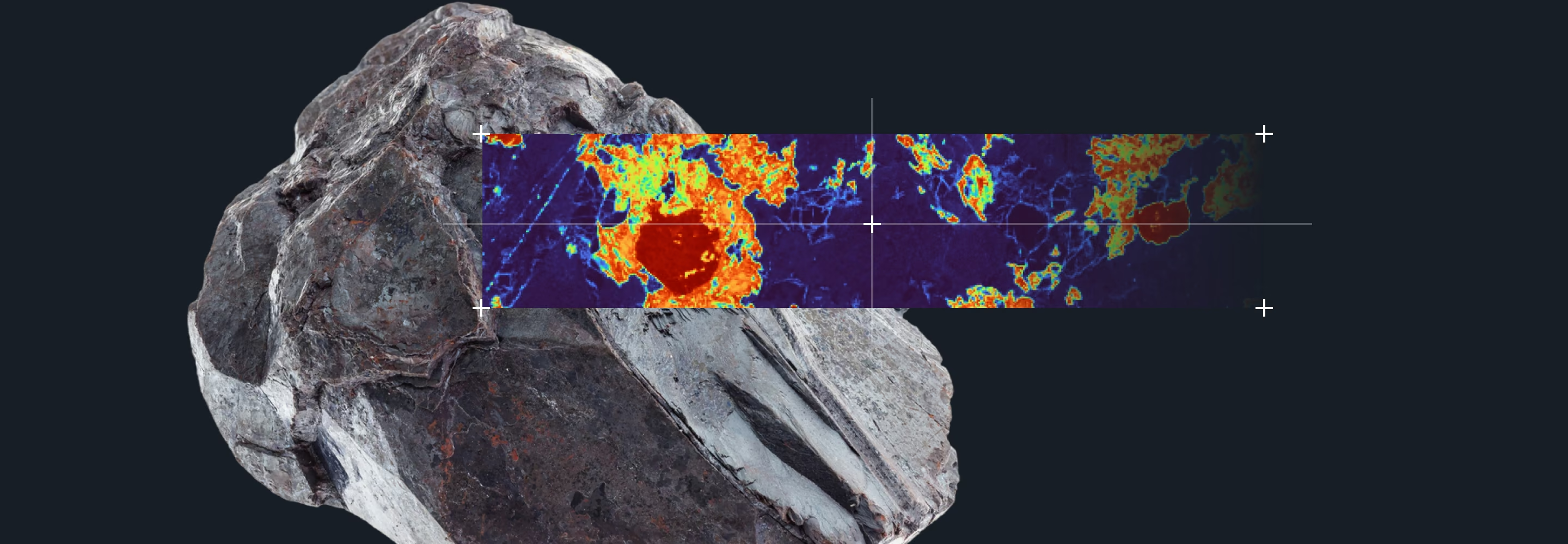
Elemental Distribution Across Battery Components
What the map shows, and which elements it's built to catch.
MicroXRF maps can reveal variations in electrode coating, current collector defects, contamination, and transition-metal crossover between cell components — spatial detail a bulk measurement can't provide.
Elements of interest, depending on chemistry:

- Electrode coating variation and thickness
- Current-collector defects
- Localized contamination
- Transition-metal crossover between cell components

Real Atlas Hardware,
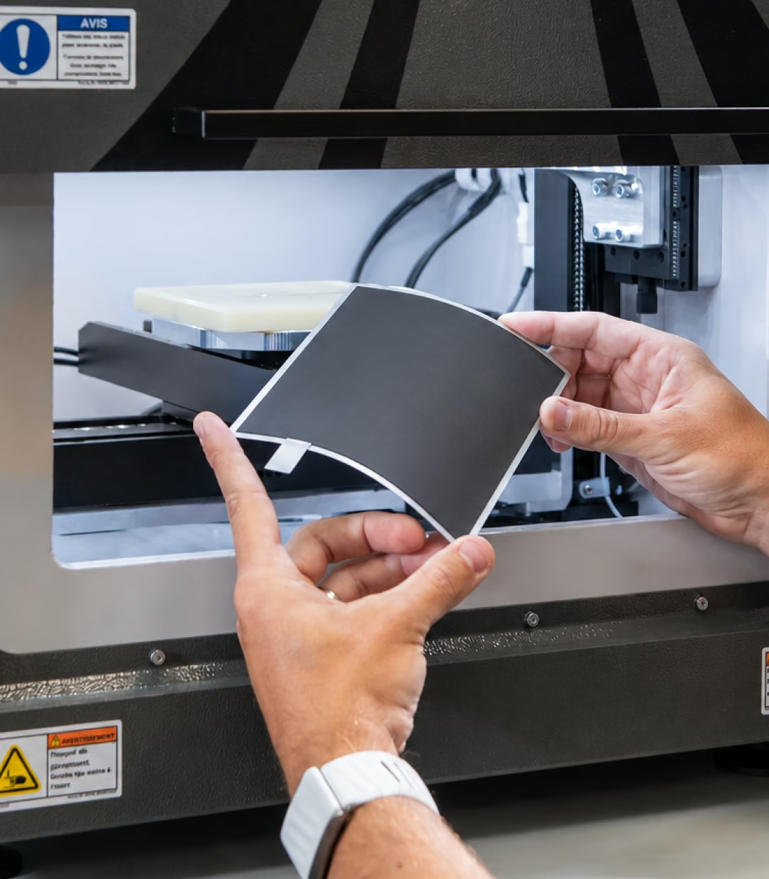
Ready for Battery Samples
The same automated chamber and sample tray used across every IXRF Atlas application.
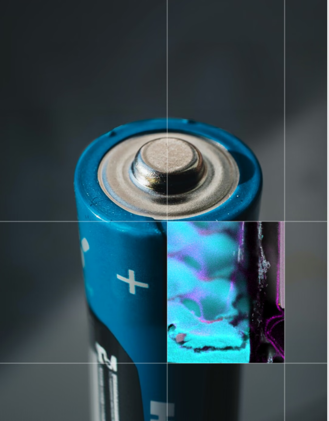
Battery Cell Cross-Section
A real elemental map, at cell scale.
This elemental map shows a primary lithium/manganese-dioxide cell in longitudinal cross-section — the wound-electrode structure, current-collector foils, and casing materials are all clearly resolved. It illustrates the kind of internal structural detail microXRF reveals at the whole-cell level, complementary to the electrode-level chemistry work described above.
How MicroXRF Analysis Works on Battery Samples
- 01.
Mount the sample — electrode coupon, cell cross-section, or current-collector foil
- 02.
Acquire an optical overview image for region-of-interest selection
- 03.
Run a focused-beam elemental map across the region of interest
- 04.
Store a full spectrum at every pixel — revisit the dataset without rescanning
- 05.
Quantify coating thickness, contamination, or crossover from the map data
MicroXRF Compared With Other Battery-Analysis Methods
MicroXRF doesn’t replace bulk or lithium-specific techniques — it fills the spatial gap between them.
vs. ICP-MS / ICP-OES
ICP methods provide sensitive bulk concentrations after the sample is dissolved. MicroXRF preserves the sample and adds the spatial distribution ICP cannot show — useful together when both bulk accuracy and location matter.
vs. SEM-EDS
SEM-EDS resolves finer features but typically requires a small, prepared, conductive-coated sample. MicroXRF covers larger electrode areas with less preparation and is often used to screen before a targeted SEM-EDS follow-up.
Important limitation:microXRF does not detect lithium directly — it falls below the technique’s typical elemental detection range. A complementary method is required for lithium-specific measurements.
See What MicroXRF Can Map in Your Battery Materials
Describe your electrode chemistry, sample geometry, and research objective to an IXRF applications scientist. We can help determine whether microXRF is appropriate and discuss sample feasibility before instrument selection.

Frequently
Asked Questions
No. Lithium emits X-rays that are too low in energy for conventional XRF detection. However, microXRF can map the transition metals, dopants, contaminants, and other elements associated with lithium-bearing battery materials, providing valuable spatial information about composition and uniformity.
MicroXRF can map many elements commonly found in battery materials, including nickel, cobalt, manganese, iron, aluminum, magnesium, phosphorus, sulfur, copper, titanium, and other metallic or inorganic constituents. Detectability depends on concentration, sample matrix, and analytical conditions.
Atlas Apex can analyze a wide range of battery-related materials, including cathodes, anodes, separators, current collectors, electrode coatings, precursor powders, active materials, cross-sections, corrosion deposits, and assembled or partially assembled components where geometry allows access to the area of interest.
Yes. MicroXRF can produce elemental maps showing how key elements are distributed across cathodes, electrodes, and coated materials. These maps can reveal composition gradients, non-uniform mixing, localized enrichment, coating variation, and other spatial heterogeneity that may affect battery performance.
MicroXRF is used to examine the spatial distribution of elements across lithium-ion battery electrodes without requiring dissolution of the sample. Researchers can use elemental maps to evaluate cathode uniformity, dopant distribution, contamination, degradation, and changes in material composition across large electrode areas.
Yes. MicroXRF is well suited for locating and identifying many types of metallic contamination, corrosion products, foreign particles, and elemental deposits on battery components. Elemental mapping can show both the chemistry and spatial distribution of anomalous material across separators, current collectors, electrodes, and other components.
In many applications, no. MicroXRF can often analyze battery materials with little or no destructive preparation, allowing electrodes, coatings, separators, and other components to be examined intact. Sectioning or polishing may still be useful when internal interfaces or specific cross-sectional features need to be investigated.
Potentially, yes. Because X-rays can penetrate through suitable windows and sample environments, microXRF can support certain in-situ or operando experimental designs when the cell geometry and materials provide sufficient X-ray access. The feasibility depends on the cell design, elements of interest, and required spatial and temporal resolution.
MicroXRF provides non-destructive elemental mapping over comparatively large areas with minimal sample preparation. SEM-EDS offers much higher spatial resolution for very small features, while ICP-MS provides highly sensitive bulk elemental measurements after sample preparation or digestion. Together, the techniques provide complementary spatial, microstructural, and quantitative chemical information.
Yes. MicroXRF can provide quantitative elemental analysis in addition to elemental mapping when appropriate calibration or Fundamental Parameters methods are used. Quantitative accuracy depends on factors such as sample matrix, surface condition, layer structure, elemental concentration, and calibration strategy.