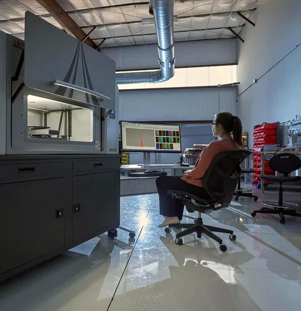
MicroXRF vs EPMA-WDS
Large-Area Elemental Mapping or High-Precision Microanalysis?
MicroXRF and electron probe microanalysis with wavelength-dispersive spectroscopy (EPMA-WDS) both provide spatially resolved elemental information, but they operate at different analytical scales.
EPMA uses a focused electron beam and wavelength-dispersive spectrometers to perform detailed quantitative analysis of small, carefully prepared regions. MicroXRF uses a focused X-ray beam to examine larger, thicker, and often minimally prepared specimens.
EPMA-WDS is generally stronger for highly localized quantitative microanalysis, light-element performance, and difficult spectral overlaps. MicroXRF is often more practical for trace-sensitive mapping across large areas, intact specimens, and samples that cannot be polished, coated, or placed under electron-beam conditions.
MicroXRF and EPMA-WDS at a Glance
When Is MicroXRF the Better Choice?
MicroXRF is particularly useful when the specimen is:
- Too large for a conventional EPMA chamber
- Too thick or irregular to prepare as a polished mount
- Nonconductive and should not be coated
- Valuable or difficult to alter
- Heterogeneous over a large area
- Being screened for trace-element-rich regions
- Part of a large mapping or survey project
- Intended for additional analytical techniques
- A complete thin section, rock slab, biological section, or manufactured component
The ability to examine intact materials can preserve relationships between grains, interfaces, inclusions, coatings, and structural features that may be changed or removed during polishing and mounting.
When Is EPMA-WDS the Better Choice?
EPMA-WDS is generally preferred when the application requires:
- Higher spatial resolution
- Precise quantitative mineral chemistry
- Measurement of very small grains or inclusions
- Better separation of overlapping X-ray peaks
- Detailed light-element analysis
- Analysis of minor elements in a carefully controlled matrix
- Correlation with high-resolution electron images
- Measurement of polished geological or metallurgical sections
- Standards-based quantitative microanalysis
Trace Sensitivity Depends on the Analytical Problem
It is overly simplistic to state that one technique is always more sensitive.
EPMA-WDS can provide excellent minor- and trace-element results when the feature is sufficiently large, the sample is properly prepared, suitable standards are available, and adequate counting time is used.
MicroXRF can provide an advantage for many mid- and higher-atomic-number elements because direct X-ray excitation typically produces lower spectral background than electron excitation. It also interrogates a larger material volume, which can improve the measured signal from distributed trace constituents.
The right technique depends on the element, matrix, feature size, concentration, and required uncertainty.
Sample Preparation and Throughput
EPMA measurements are normally performed on flat, polished, vacuum-compatible specimens. Nonconductive materials commonly require a conductive coating.
These requirements provide controlled analytical geometry but can add preparation time and may make EPMA unsuitable for:
- Rough surfaces
- Large objects
- Delicate materials
- Wet or volatile samples
- Objects that cannot be sectioned
- Samples whose original surface must be preserved
MicroXRF can often begin with minimal sample preparation.
A Complementary Workflow
MicroXRF and EPMA-WDS can form an efficient multiscale workflow:
- Use microXRF to map the complete specimen.
- Locate trace-element enrichments, zoning, inclusions, and chemically distinct regions.
- Select representative areas for sectioning or polishing.
- Use EPMA-WDS for precise mineral chemistry and higher-resolution analysis.
- Correlate the localized EPMA results with the broader elemental map.
This can reduce time spent searching for small, analytically important features using the electron probe.
Why Atlas Apex for Large-Area Microanalysis?
Atlas Apex provides:
- X-ray spot sizes down to approximately 5 µm
- Large automated mapping areas
- Perpendicular excitation geometry
- Multiple silicon drift detector configurations
- Full-spectrum data retained at every map pixel
- Air, helium, and vacuum operation
- Quantitative mapping and spot-analysis workflows
- Statistical clustering and phase-segmentation tools
- Accommodation of large and irregular specimens