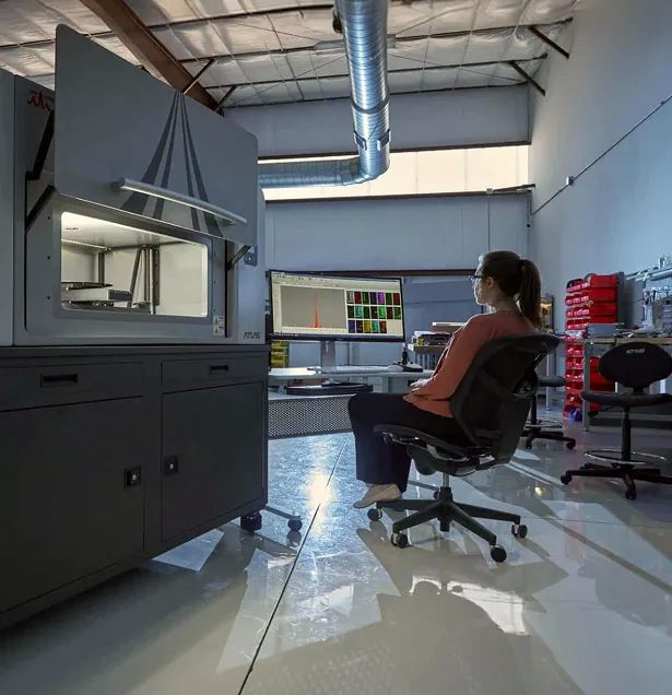
MicroXRF vs Raman Microscopy
Elemental Composition or Molecular Identity?
MicroXRF and Raman microscopy can both create detailed chemical maps, but they measure fundamentally different properties.
MicroXRF detects characteristic X-rays to determine which elements are present and where they are distributed.
Raman microscopy measures how laser light interacts with molecular bonds and crystal structures. It can identify compounds, minerals, pigments, polymers, and other materials based on their molecular or structural fingerprints.
MicroXRF answers:
Which elements are present, and where are they located?
Raman microscopy answers:
Which compounds or molecular structures are present?
MicroXRF and Raman Microscopy at a Glance
When Is MicroXRF the Better Choice?
MicroXRF is preferred when the primary question concerns elemental composition.
Examples include:
- Mapping metals within biological tissue
- Locating heavy-metal contamination
- Measuring elemental zoning in minerals
- Finding rare-earth-element or critical-mineral enrichments
- Examining alloy segregation
- Characterizing metallic pigments and inorganic fillers
- Mapping corrosion, diffusion, or reaction fronts
- Identifying elemental associations within inclusions
- Analyzing areas larger than are practical for detailed Raman mapping
- Investigating materials affected by optical fluorescence
MicroXRF can detect an element even when it is present in multiple chemical or structural forms. This makes it especially useful for locating the complete distribution of a metal or other analyte before determining its specific compound or phase.
When Is Raman Microscopy the Better Choice?
Raman microscopy is the stronger technique when the analysis requires:
- Molecular identification
- Mineral-phase confirmation
- Polymer identification
- Differentiation of carbon allotropes
- Identification of organic compounds
- Pharmaceutical polymorph analysis
- Pigment or dye characterization
- Chemical-bonding information
- Identification of compounds with identical elemental compositions
- High-spatial-resolution examination of small particles or defects
For example, microXRF may show that two regions contain similar elements. Raman microscopy may determine that those elements are arranged in different molecular or crystalline structures.
Elements Are Not the Same as Compounds
An elemental map does not always identify the material responsible for the signal.
A region containing iron could represent:
- Metallic iron
- An iron oxide
- An iron sulfide
- An iron-bearing silicate
- An iron-containing pigment
- A dispersed contaminant
MicroXRF can show the iron distribution and its association with other elements. Raman microscopy may identify the specific compound or mineral phase.
The reverse is also true: Raman may identify a compound while providing limited information about low-concentration elemental impurities or trace-metal distributions.
Important Raman Considerations
Raman analysis can be affected by:
- Fluorescence that overwhelms the Raman signal
- Laser heating
- Burning or alteration of sensitive materials
- Surface roughness
- Crystal orientation
- Weak scattering from some compounds
- Long acquisition times for large, high-resolution maps
These factors determine which samples and analytical questions are best matched to Raman microscopy.
A Complementary Workflow
MicroXRF and Raman microscopy are highly complementary:
- Use microXRF to map elemental distributions across the complete sample.
- Identify chemically distinct regions and elemental associations.
- Select representative points or small regions.
- Use Raman microscopy to identify molecular compounds or mineral phases.
- Correlate molecular identity with the broader elemental map.
This workflow is particularly valuable for geology, battery research, cultural heritage, polymers, environmental particles, pharmaceuticals, and biological materials.
Why Atlas Apex for Elemental Context?
Atlas Apex provides:
- X-ray spot sizes down to approximately 5 µm
- Large-area elemental mapping
- Full-spectrum data at every pixel
- Trace-sensitive analysis of many metallic and inorganic elements
- Quantitative spot analysis and mapping
- Air, helium, and vacuum operation
- Automated linescans and region analysis
- Element-ratio imaging
- Statistical clustering of chemically similar pixels
- Repeatable return to selected locations for complementary analysis