Company News
Explore practical resources covering microXRF, XRF analysis, elemental mapping, instrumentation, and real-world applications.
This learning center is designed for researchers, laboratory managers, engineers, educators, and technical buyers who want to better understand microXRF and its role in elemental analysis.
Build your understanding of microXRF through detailed guides, technical explainers, and step-by-step educational content — how XRF works, sample preparation, elemental mapping, quantitative analysis, instrument selection, and application-specific workflows.
Compare microXRF with other elemental and materials-analysis techniques to determine which approach best fits your sample and analytical objective — sample prep, spatial resolution, elemental sensitivity, mapping capability, and workflow.

MicroXRF, or micro X-ray fluorescence spectroscopy, is a non-destructive elemental analysis technique that uses a focused X-ray beam to measure the chemical composition of small areas on a sample. It can analyze individual points, lines, or large areas to reveal how elements are distributed spatially.
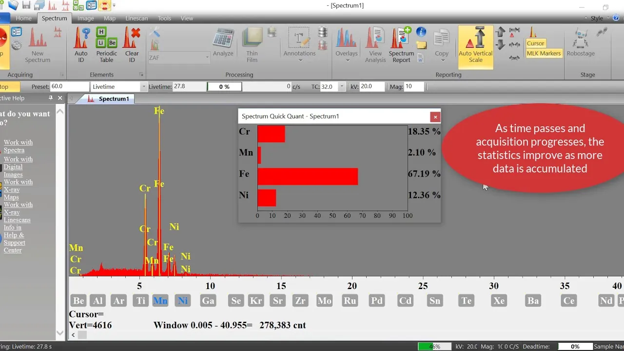
A focused X-ray beam excites atoms in the sample, causing them to emit characteristic fluorescent X-rays. An energy-dispersive detector measures these X-rays, and the resulting spectrum identifies the elements present and provides information about their relative or quantitative concentrations.
Conventional XRF typically measures the average composition of a relatively large sample area, while microXRF uses a much smaller beam to provide spatially resolved elemental information. This allows microXRF to identify localized inclusions, phases, coatings, gradients, contamination, and other chemical variations that may be hidden in a bulk measurement.
MicroXRF uses X-rays for excitation, while SEM-EDS uses an electron beam. SEM-EDS generally provides much higher spatial resolution, while microXRF can analyze larger samples and areas, offers greater X-ray penetration, and often provides stronger sensitivity for trace and higher-energy elements with minimal sample preparation.
The detectable elemental range depends on the X-ray source, detector, window configuration, atmosphere, sample matrix, and concentration. Modern microXRF systems can analyze a broad range of elements, with specialized configurations extending detection from light elements such as carbon or fluorine through uranium. Lithium cannot be measured directly by conventional XRF.
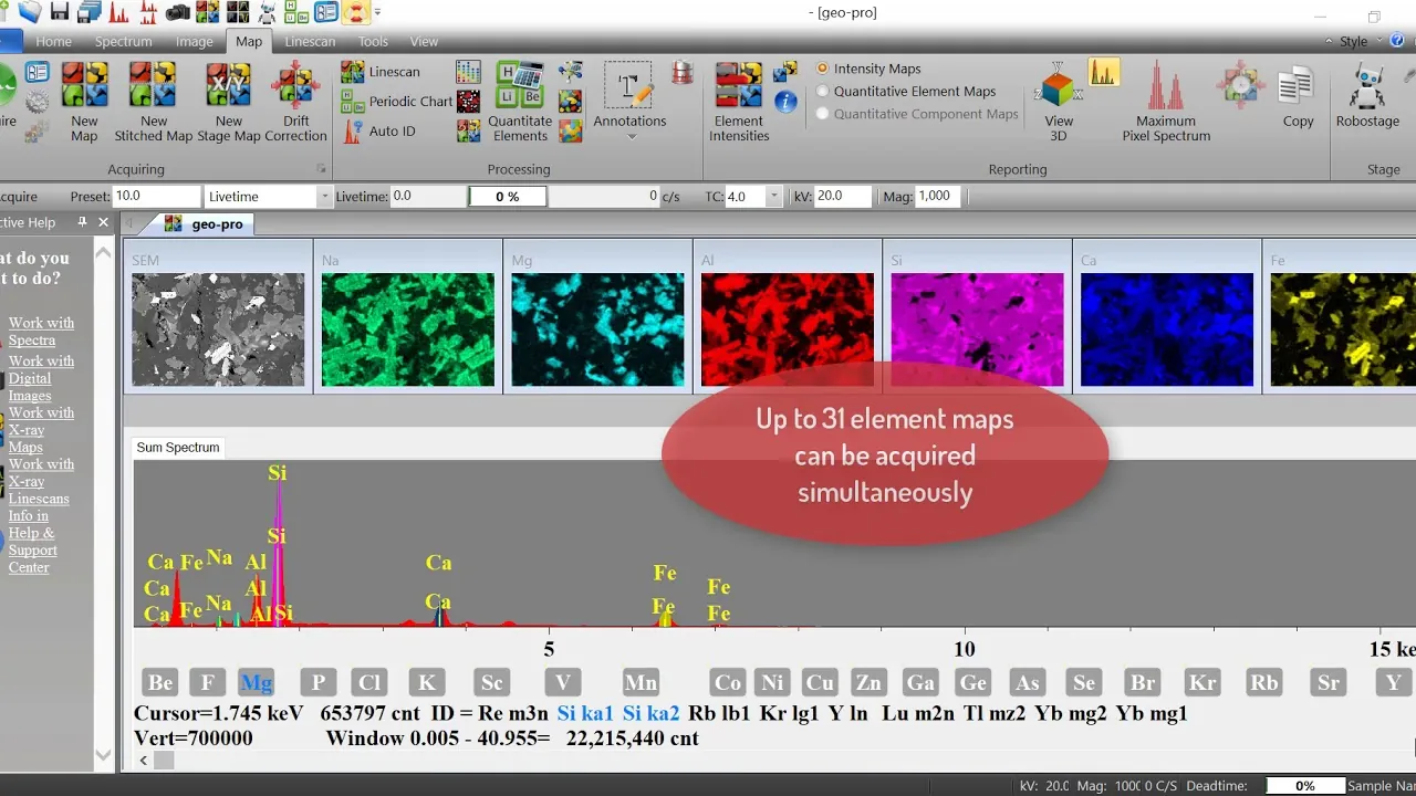
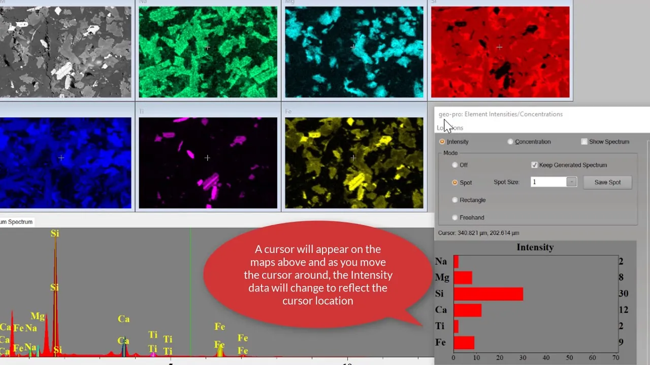
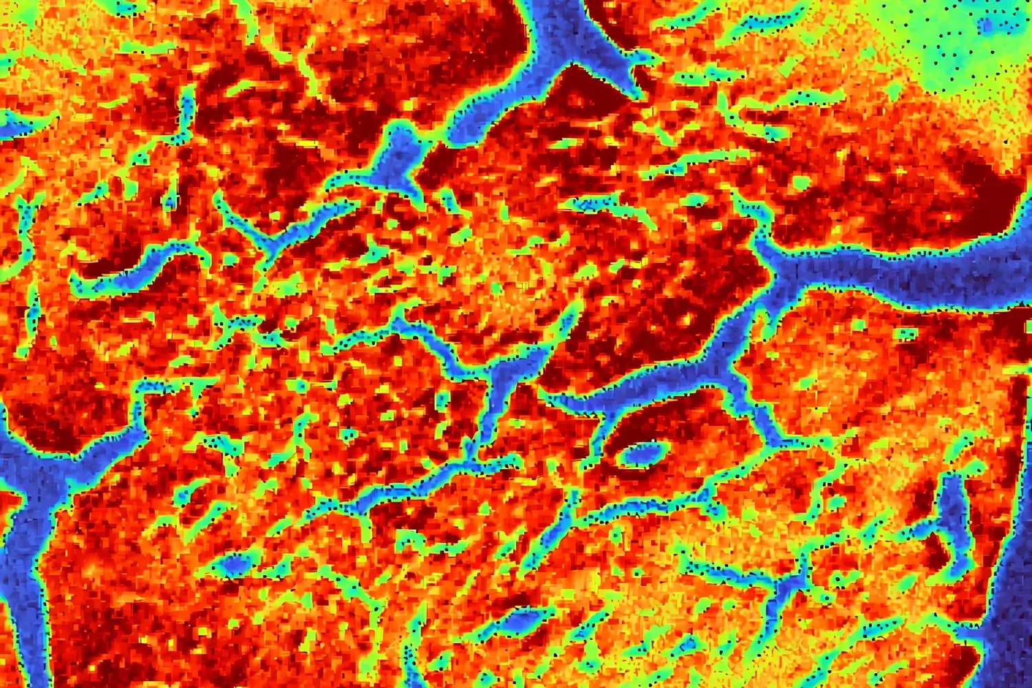
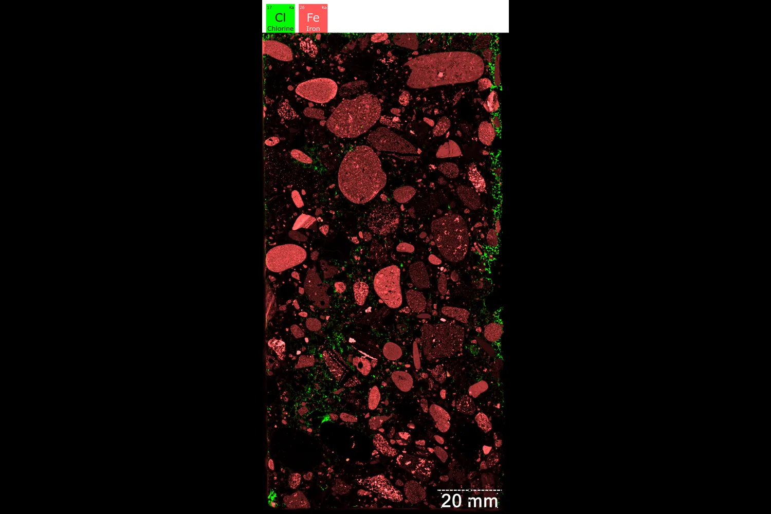
An elemental map is a two-dimensional image showing the spatial distribution of one or more elements across a sample. The instrument collects an X-ray spectrum at each measurement position, allowing individual element maps and chemical relationships to be generated from the same dataset.
Spot size describes the approximate diameter of the focused X-ray beam at the sample. Spatial resolution describes how closely two chemically different features can actually be distinguished and is also influenced by X-ray penetration, fluorescence within the sample, elemental energy, sample composition, surface geometry, and measurement conditions.
Yes. MicroXRF can provide quantitative concentration data using calibration standards, Fundamental Parameters methods, or other quantitative approaches. Accuracy depends on factors including sample matrix, surface condition, thickness, homogeneity, calibration quality, and the elements being measured.
Often very little. Many solids, geological specimens, electronic components, artifacts, and other materials can be analyzed directly. However, flat, clean, homogeneous, and polished surfaces generally improve quantitative accuracy and spatial resolution when precise measurements are required.
Air absorbs low-energy X-rays, reducing sensitivity to lighter elements. Vacuum removes the air path, while helium displaces air with a gas that absorbs far fewer low-energy X-rays. Both approaches can significantly improve light-element detection, with helium particularly useful for samples that should not be placed under vacuum.
Detection limits depend on the element, sample matrix, X-ray source intensity, excitation conditions, detector efficiency, acquisition time, background level, spot size, atmosphere, and sample geometry. Longer measurement times and optimized analytical conditions generally improve sensitivity.
MicroXRF is particularly useful when researchers need non-destructive elemental analysis combined with spatial information. Applications include geology and critical minerals, metals, batteries, semiconductors and microelectronics, ceramics, concrete, environmental materials, cultural heritage, coatings, forensic samples, and advanced materials research.