Table of contents
Laboratory-Based Elemental Imaging Reveals Phosphorus, Iron, and Copper Distributions in Human Brain Tissue
Alzheimer’s disease is characterized by complex biochemical and structural changes within brain tissue. Understanding those changes requires analytical methods capable of connecting molecular and elemental information with the microscopic pathological features observed by neuropathologists.
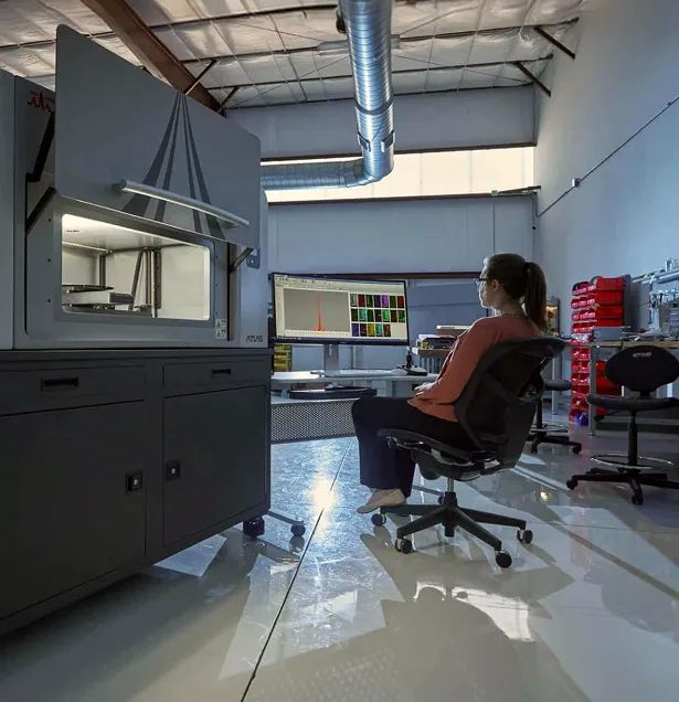
In collaboration with Prof. Lee Makowski and his research team at Northeastern University, IXRF Systems used laboratory-based microXRF to investigate elemental distributions within histological thin sections of human brain tissue affected by Alzheimer’s disease.
The Atlas microXRF mapped phosphorus (P), iron (Fe), and copper (Cu) with micron-scale spatial resolution. The resulting elemental maps were compared with serial tissue sections stained for pathological features including tau and amyloid-β.
The study demonstrated how laboratory microXRF can add spatially resolved elemental information to established histopathology workflows without altering the morphology of the analyzed tissue.
The Challenge
Connecting Elemental Chemistry With Alzheimer’s Pathology
The defining pathological features of Alzheimer’s disease include amyloid-β plaques and abnormal accumulations of tau protein.
Traditional histological staining is essential for locating these structures, but staining alone does not provide direct information about how chemical elements are distributed around them.
Researchers therefore wanted to investigate whether elemental changes within the tissue corresponded spatially with neuropathological features.
Of particular interest was phosphorus.
Phosphorylation of tau is closely associated with Alzheimer’s pathology, making localized phosphorus distribution an important subject of investigation. Iron and copper were also examined because their distributions could provide additional information about the chemical environment surrounding diseased tissue.
The analytical challenge was therefore not simply to determine whether these elements were present.
Researchers needed to determine:
Where are phosphorus, iron, and copper located relative to pathological structures within the tissue?
Why Spatial Information Matters
A bulk measurement of brain tissue can provide an average elemental concentration, but that average eliminates the spatial relationships between chemistry and tissue structure.
This becomes particularly important when the structures of interest occupy only small regions within a much larger tissue section.
Spatially resolved elemental analysis allows researchers to investigate questions such as:
- Is an element enriched near a pathological feature?
- Does elemental concentration change across neighboring tissue regions?
- Do different pathological structures exhibit different elemental signatures?
- Can elemental maps be correlated with conventional histological staining?
MicroXRF provides a way to address these questions while maintaining the spatial context of the original tissue.
The Analytical Approach
Mapping Histological Thin Sections With MicroXRF
The research team analyzed histological thin sections of human brain tissue using the Atlas microXRF.
A focused X-ray beam was scanned across selected tissue regions while energy-dispersive detectors collected characteristic X-rays emitted by the sample.
From those measurements, researchers generated spatial maps showing the distributions of:
- Phosphorus
- Iron
- Copper
The Atlas system provided approximately 5 µm-scale spatial capability for the elemental mapping performed in the collaboration.
Because XRF analysis does not require conventional chemical staining of the analyzed section, elemental information could be collected while retaining the morphology of the specimen.
Correlating MicroXRF With Histopathology
Using Serial Sections to Connect Chemistry and Structure
One of the most important aspects of the study was the comparison of microXRF data with conventional neuropathological observations.
Serial tissue sections were stained for:
- Tau
- Amyloid-β
These stained sections were compared with the elemental maps obtained from nearby sections.
This allowed researchers to examine whether localized elemental distributions corresponded with recognized pathological features.
The approach combines two complementary forms of information:
- Histopathology identifies the biological structures.
- MicroXRF identifies the elemental distributions associated with those regions.
Together, the two techniques provide a more complete picture than either measurement alone.
The Results
1. Phosphorus Enrichment Was Observed Near Pathological Features
The most significant observation was increased phosphorus signal in regions adjacent to tau fibrils.
This spatial enrichment was consistent with observations discussed in previous synchrotron-based studies of pathological protein-rich regions.
Rather than detecting only the total amount of phosphorus within the tissue section, microXRF showed where phosphorus was concentrated relative to disease-associated structures.
This illustrates an important advantage of elemental mapping in neuropathology: potentially significant biochemical variations can be examined while preserving their spatial relationship to tissue morphology.
2. Iron and Copper Showed Additional Spatial Patterns
The study also mapped iron and copper.
The original investigation describes these observations as preliminary relative to the phosphorus findings, but both elements displayed spatial distributions that could warrant further investigation.
Iron and copper mapping may provide complementary information about the elemental environment surrounding pathological structures.
Importantly, the study does not establish a causal role for these elemental patterns in Alzheimer’s disease. Instead, the maps identify spatial relationships that can guide additional investigation.
3. Elemental Maps Could Be Compared With Tau and Amyloid-β Staining
Comparing microXRF results with serial sections stained for tau and amyloid-β provided an important validation strategy.
Instead of interpreting elemental patterns in isolation, researchers could examine those patterns alongside established neuropathological markers.
This correlation enabled researchers to connect:
Elemental distribution → tissue location → pathological structure
That layered analytical approach is one of the strongest aspects of the workflow.
Why Laboratory-Based MicroXRF Matters
Moving Elemental Imaging Closer to the Research Laboratory
High-resolution X-ray fluorescence microscopy has traditionally been associated with synchrotron facilities.
Synchrotrons provide exceptional analytical capabilities, but they also involve practical limitations:
- Competitive beam-time allocation
- Travel to specialized facilities
- Limited experimental schedules
- Complex logistics
- Restricted opportunities for routine or iterative measurements
Laboratory microXRF does not replace synchrotron XRF microscopy.
Instead, it provides researchers with an accessible complementary capability for experiments that can be performed at the spatial resolution and sensitivity available from a laboratory instrument.
This can enable researchers to perform routine elemental mapping, refine experimental methods, screen samples, and identify high-value specimens or regions before pursuing more specialized measurements.
Preserving Valuable Biological Samples
Elemental Information Without Traditional Destructive Preparation
Biological specimens used in neurological research can be limited and scientifically valuable.
MicroXRF provides elemental information without requiring the tissue to be dissolved or consumed during analysis.
This allows the spatial arrangement of the sample to be preserved and facilitates correlation with complementary analytical methods.
For neuropathology research, preserving spatial context is particularly important because the relationship between a chemical signal and a specific tissue structure may provide more useful information than the average chemical composition of the entire specimen.
A Multimodal Research Workflow
The collaboration illustrates how microXRF can fit into a broader biological imaging workflow.
1. Prepare Histological Sections
Generate tissue sections suitable for elemental analysis and complementary histopathological evaluation.
2. Map the Tissue With MicroXRF
Collect spatially resolved elemental maps of selected regions.
3. Identify Elemental Features
Examine phosphorus, iron, copper, or other detectable elemental distributions for localized enrichment or depletion.
4. Compare Serial Sections
Correlate the elemental maps with adjacent sections stained for relevant pathological markers.
5. Identify Regions of Interest
Locate areas where elemental distributions correspond with neuropathological structures.
6. Apply Complementary Techniques
Use those regions to guide additional microscopy, spectroscopy, synchrotron analysis, or other specialized investigations where appropriate.
This workflow allows microXRF to act as a bridge between traditional histology and advanced chemical imaging.
From Phosphorus Maps to New Research Questions
The significance of the experiment is not that microXRF alone explains Alzheimer’s disease.
Its value is in enabling researchers to ask more spatially specific biochemical questions.
For example:
- How consistently does phosphorus enrichment occur near tau-rich regions?
- Do elemental distributions vary between different pathological structures?
- How do elemental signatures differ between affected and comparison tissues?
- Are iron or copper distributions consistently associated with particular tissue features?
- Can elemental patterns be correlated with disease stage or other neuropathological measurements?
These questions require larger, controlled studies before conclusions can be drawn.
MicroXRF provides an analytical platform capable of contributing spatial elemental data to those investigations.
The Value
Adding Chemical Context to Histopathology
This case study demonstrates several advantages of laboratory microXRF for biological tissue research.
Spatially Resolved Elemental Information
Researchers can visualize where selected elements occur rather than relying exclusively on bulk concentration measurements.
Correlation With Histological Features
Elemental maps can be compared with serial sections stained using established neuropathological methods.
Minimal Sample Alteration
The technique preserves the morphology of the analyzed specimen.
Laboratory Accessibility
Researchers can perform microXRF measurements without relying exclusively on synchrotron access.
Broad Elemental Mapping
Multiple elements can be investigated from the same XRF dataset, allowing researchers to examine relationships among elemental distributions.
Together, these capabilities make laboratory microXRF a useful complementary tool for research questions where elemental chemistry and tissue structure need to be studied together.
The Takeaway
Connecting Elemental Chemistry With Alzheimer’s Pathology
Understanding neurodegenerative disease requires analytical techniques capable of examining complex biological processes at multiple scales.
In this collaboration with Prof. Lee Makowski’s team at Northeastern University, laboratory-based microXRF was used to map phosphorus, iron, and copper in histological sections of human brain tissue affected by Alzheimer’s disease.
The study identified phosphorus enrichment near pathological features and demonstrated the value of comparing spatial elemental maps with serial sections stained for tau and amyloid-β.
The result is not a diagnostic test or proof of a specific disease mechanism.
It is a demonstration of a powerful research workflow: combining elemental imaging with conventional neuropathology to investigate where chemical changes occur relative to disease-associated structures.
For neuroscience, neuropathology, and other biological research disciplines, laboratory microXRF provides a new way to add elemental context to microscopic tissue analysis.
Explore MicroXRF for Biological and Life Science Research
Interested in investigating metals, minerals, nutrients, or other elemental distributions within biological samples?
See how Atlas Apex microXRF can add spatially resolved elemental information to your research workflow.