Table of contents
Using 5 µm Elemental Mapping to Identify Regions of Interest in Historical FFPE Lung Tissue
Synchrotron X-ray fluorescence microscopy can provide exceptional spatial resolution and elemental sensitivity for biomedical research. But access to synchrotron facilities is limited, beam time is competitive, and researchers may have only a short window to analyze valuable specimens.
That creates an important practical question:
Can laboratory microXRF perform the initial elemental mapping needed to identify the most important regions before a sample reaches the synchrotron?
Working with Tatjana Paunesku and Jeffrey Murley of Northwestern University, IXRF Systems evaluated this question using archival formalin-fixed, paraffin-embedded (FFPE) lung tissue that had previously been characterized using synchrotron X-ray fluorescence microscopy.
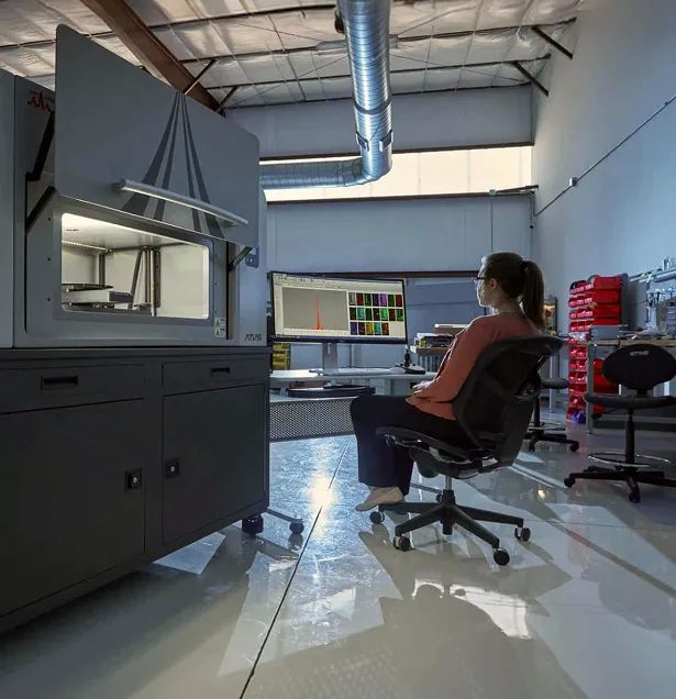
The tissue originated from a historical radiobiology study involving a beagle exposed to yttrium-90-loaded aluminosilicate microparticles. Using an Atlas X microXRF equipped with a 5 µm focused X-ray beam, the laboratory system mapped silicon, phosphorus, sulfur, calcium, and iron and successfully identified silicon-rich regions suitable for closer investigation.
The comparison demonstrated a practical role for laboratory microXRF as a screening and region-of-interest selection tool that complements higher-resolution synchrotron analysis.
The Challenge
Valuable Biological Samples, Limited Synchrotron Access
Synchrotron XFM has become an important technique for studying elemental distributions within biological tissues.
Its combination of sensitivity and very high spatial resolution allows researchers to investigate elemental distributions at cellular and, in some cases, subcellular scales.
However, synchrotron access creates practical limitations.
Researchers generally must:
- Submit proposals for beam time
- Compete for limited facility availability
- Transport samples and experimental materials
- Conduct experiments within fixed scheduling windows
- Decide in advance which samples and regions deserve detailed analysis
For rare or archival biological specimens, these limitations make experiment planning particularly important.
Using valuable beam time to search a large tissue section for a few relevant microscopic regions is not always the most efficient workflow.
A laboratory technique capable of screening larger areas first could allow researchers to arrive at the synchrotron already knowing where to focus.
The Biological Sample
Archival FFPE Tissue From a Historical Radiation Study
The tissue examined in this case originated from a historical radiobiology experiment conducted in 1968.
Beagle dogs were exposed by inhalation to insoluble aluminosilicate microparticles carrying the beta-emitting radionuclide yttrium-90 (^90Y).
Many of the resulting formalin-fixed, paraffin-embedded specimens were preserved and are now part of the Northwestern University Radiobiology Archive.
Because ^90Y has a short half-life, the original radionuclide was no longer present when the samples were examined decades later. However, the aluminosilicate carrier particles and elemental signatures associated with them remained available for investigation.
The published synchrotron study used these archival specimens to determine whether XFM could locate formerly radioactive microparticles and characterize their elemental environment decades after the original exposure.
The Original Synchrotron Study
Mapping Radioactive-Particle History Through Elemental Signatures
The original study analyzed FFPE lung and lymph-node tissue using several XFM beamlines at the Advanced Photon Source at Argonne National Laboratory.
Researchers mapped elements including:
- Silicon
- Phosphorus
- Sulfur
- Iron
- Zinc
Silicon served as an important marker because it was a component of the aluminosilicate particles used to carry ^90Y.
The synchrotron measurements identified silicon-rich microparticles in both lung and lymph-node tissue. Researchers also observed associations between silicon and iron at lower and intermediate spatial resolutions.
At higher resolution, the relationship became more complex: iron signals were often located adjacent to, rather than precisely overlapping, individual silicon-rich particles.
The original study also detected zirconium, the stable decay product of ^90Y, associated with silicon-rich regions in lymph-node tissue. That result supported the conclusion that the microparticles remained sufficiently intact during cellular transport to retain evidence of their radioactive history.
The Question for Laboratory MicroXRF
The synchrotron results established what could be accomplished with advanced XFM.
IXRF and the Northwestern researchers then addressed a different question:
How much of the preliminary elemental mapping could be performed with a laboratory microXRF before using synchrotron beam time?
The goal was not to reproduce nanoscale synchrotron performance.
Instead, the objective was to determine whether laboratory microXRF could:
- Survey a larger biological tissue area
- Identify silicon-rich regions
- Map major elemental distributions
- Locate regions deserving higher-resolution analysis
- Reduce the amount of exploratory work required at a synchrotron
The Analytical Approach
Reanalyzing the Tissue With Laboratory MicroXRF
A tissue sample from the original investigation was analyzed using the Atlas X microXRF.
The system was equipped with a polycapillary-focused X-ray source providing a 5 µm X-ray spot size.
Elemental maps were collected for:
- Si — Silicon
- P — Phosphorus
- S — Sulfur
- Ca — Calcium
- Fe — Iron
The maps provided an overview of elemental distributions throughout the analyzed tissue while maintaining substantially finer spatial information than conventional bulk XRF analysis.
Researchers then generated merged elemental maps and selected smaller regions for additional sub-mapping.
This created a hierarchical workflow:
Large-area survey → identify elemental features → select region of interest → collect a more detailed map
That workflow is especially useful when the ultimate goal is to guide subsequent higher-resolution analysis.
The Results
1. Laboratory MicroXRF Identified Silicon-Rich Regions
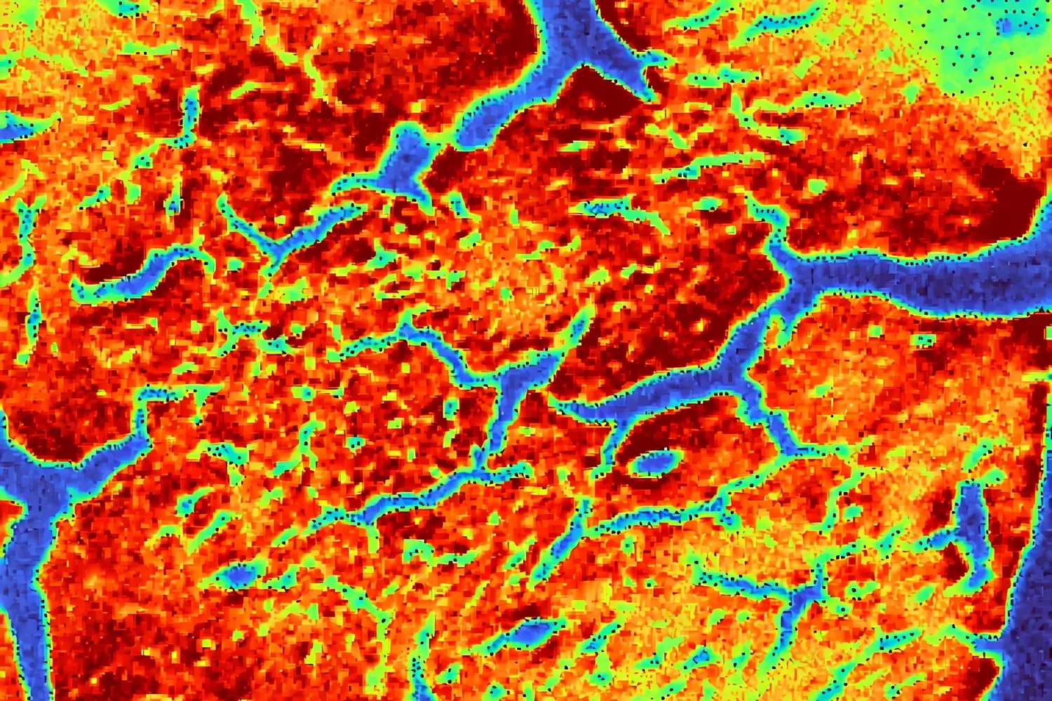
The Atlas X elemental maps clearly revealed regions enriched in silicon.
Because silicon was associated with the aluminosilicate particles used in the original exposure experiment, these regions were of particular interest.
The laboratory system therefore provided researchers with the information needed to locate candidate particle-rich areas without first using valuable synchrotron beam time to search across the specimen.
2. Multiple Tissue-Related Elements Were Mapped Simultaneously
In addition to silicon, the microXRF analysis mapped phosphorus, sulfur, calcium, and iron.
These elements provided additional chemical context around the silicon-rich regions.
The resulting maps demonstrated that laboratory microXRF could provide a broader elemental overview of the tissue while researchers searched for the features most relevant to the radiation-exposure investigation.
3. Merged Elemental Maps Added Spatial Context
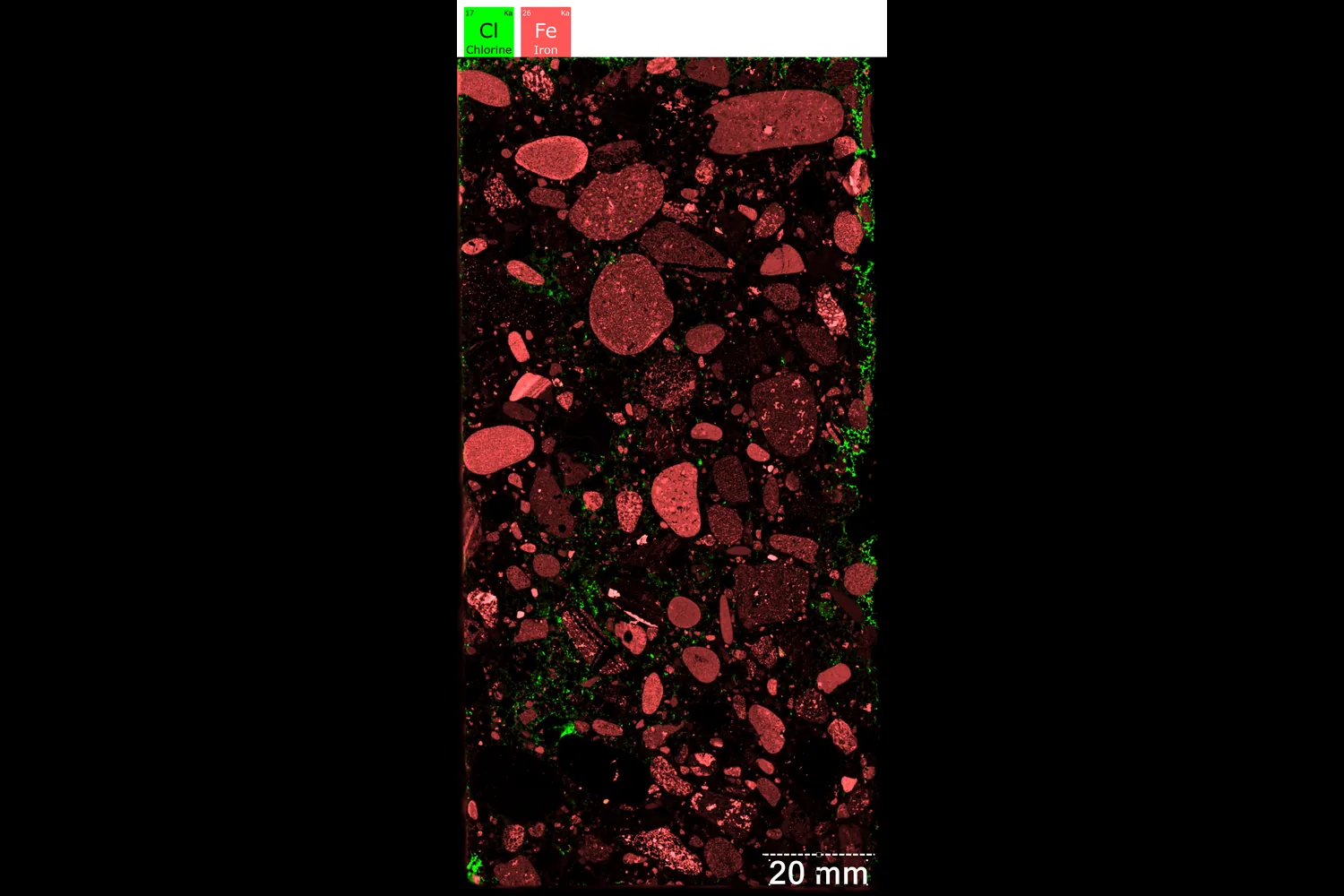
Overlaying elemental distributions allowed researchers to compare where different elemental signals occurred relative to one another.
Rather than examining an isolated silicon map, the merged data provided a more complete view of the local chemical environment.
This helped distinguish regions that warranted closer inspection and supported selection of specific areas for subsequent sub-mapping.
4. Higher-Detail Sub-Maps Could Be Collected From Selected Regions
Once a region of interest was identified, the laboratory microXRF could collect a smaller map focused on that area.
This survey-to-detail workflow is one of the most important outcomes of the case study.
Instead of acquiring maximum-detail data across an entire tissue specimen, researchers can use a broader map to locate the most informative regions and then concentrate measurement time where it matters.
The same logic can be applied when preparing samples for synchrotron analysis.
Laboratory MicroXRF vs. Synchrotron XFM
Complementary Tools at Different Spatial Scales
The case study does not demonstrate that laboratory microXRF replaces synchrotron XFM.
The synchrotron systems used in the original research provided substantially finer spatial resolution. For example, the published study used XFM measurements ranging from tens of micrometers for overview scans down to sub-micron and nanoscale beam sizes for detailed investigation.
Laboratory microXRF occupies a different analytical space.
Laboratory MicroXRF Is Well Suited For
- Large-area elemental surveys
- Sample screening
- Identifying regions of interest
- Routine experiments
- Method development
- Iterative measurements
- Preliminary analysis before synchrotron experiments
Synchrotron XFM Is Well Suited For
- Sub-micron analysis
- Cellular and subcellular elemental mapping
- Very high sensitivity
- Detailed interrogation of selected microscopic features
- Experiments requiring specialized beam energies or synchrotron capabilities
The two techniques therefore form a natural analytical sequence.
A More Efficient Synchrotron Workflow
Use Laboratory MicroXRF Before Beam Time
A research group planning a synchrotron experiment could apply the workflow demonstrated here in several stages.
1. Screen Samples In-House
Use laboratory microXRF to map candidate specimens and determine which contain relevant elemental features.
2. Select the Best Specimens
Exclude samples that do not contain the chemistry or structures required for the research question.
3. Identify Regions of Interest
Locate particle-rich, metal-rich, or otherwise chemically distinct regions before synchrotron analysis.
4. Optimize Experimental Conditions
Use laboratory measurements to determine which elements and regions deserve priority.
5. Conduct Targeted Synchrotron Analysis
Use scarce beam time for the specific regions requiring higher spatial resolution or sensitivity.
6. Return to the Laboratory for Follow-Up
Perform additional routine mapping or comparative measurements without waiting for another synchrotron allocation.
This approach can make synchrotron experiments more focused and potentially increase the scientific value obtained from each beam-time allocation.
Why This Matters for Biomedical Research
Many Biomedical Questions Depend on Spatial Chemistry
Biological tissues are chemically heterogeneous.
An averaged elemental concentration can obscure small but potentially significant regions where metals, minerals, particles, or other elements accumulate.
Spatial elemental imaging can contribute to research involving:
- Metal accumulation in tissues
- Environmental exposure
- Toxicology
- Nanoparticle distribution
- Radiation biology
- Tumor biology
- Neurodegenerative disease
- Nutrient and trace-element localization
- Drug or therapeutic-material distribution
For these applications, the location of an element may be as important as its overall concentration.
Preserving Archival and Valuable Samples
The tissue in this study also illustrates another advantage of X-ray fluorescence analysis.
FFPE archives can contain specimens that are decades old, rare, and impossible to reproduce.
The published synchrotron research demonstrated that XFM could obtain elemental information from approximately 35-year-old archival tissue specimens while preserving them for additional analysis.
Laboratory microXRF similarly provides a non-destructive approach to elemental mapping, making it particularly valuable for preliminary screening of archival collections where sample preservation is important.
Accessibility Changes the Research Workflow
A laboratory instrument provides something a synchrotron cannot easily provide:
routine access.
Researchers can perform measurements when needed rather than building every experiment around an allocated beam-time window.
This makes it possible to:
- Test more samples
- Repeat measurements
- Modify preparation methods
- Optimize scan conditions
- Train students
- Develop new analytical methods
- Conduct feasibility studies
- Respond immediately to unexpected results
That flexibility does not replace the capabilities of a synchrotron.
It helps researchers use those specialized capabilities more strategically.
The Value
Turning Synchrotron Preparation Into an Analytical Workflow
The Atlas X analysis demonstrated that a 5 µm laboratory microXRF could perform a significant portion of the initial elemental mapping and region-of-interest identification required for this biological tissue investigation.
The practical benefits include:
Better Sample Selection
Researchers can identify which samples contain relevant elemental features before committing synchrotron resources.
More Targeted Beam Time
Regions of interest can be defined before arrival at the facility.
Faster Experimental Iteration
Laboratory measurements can be repeated or modified without waiting for another beam-time allocation.
Preservation of Valuable Specimens
Elemental maps can be collected non-destructively from rare archival tissue.
Complementary Multi-Scale Analysis
Laboratory and synchrotron XRF can be combined to investigate the same scientific question across progressively finer spatial scales.
The Takeaway
Laboratory MicroXRF Does Not Need to Replace the Synchrotron to Transform the Workflow
The original synchrotron study demonstrated the remarkable elemental information that can be recovered from archival FFPE tissue, including the identification of formerly radioactive aluminosilicate microparticles and their elemental environment.
The follow-up laboratory microXRF analysis answered a different but equally practical question.
Using a 5 µm focused X-ray beam, Atlas X successfully mapped silicon, phosphorus, sulfur, calcium, and iron and identified silicon-rich regions for additional investigation.
The result demonstrates a compelling model for biomedical research:
Use laboratory microXRF to survey, screen, and select. Use synchrotron XFM when the scientific question demands the next level of spatial resolution or sensitivity.
For laboratories working with biological tissues, archival specimens, nanoparticles, environmental exposures, or other spatially complex samples, this combination can make advanced elemental imaging more accessible while helping researchers use limited synchrotron resources more efficiently.
About the Research
Laboratory MicroXRF Collaboration
IXRF Systems
Tatjana Paunesku and Jeffrey Murley, Northwestern University
Related Published Study
Proof of principle study: synchrotron X-ray fluorescence microscopy for identification of previously radioactive microparticles and elemental mapping of FFPE tissues
Journal: Scientific Reports
Published: May 13, 2023
The published study investigated archival canine lung and lymph-node tissue from a historical ^90Y-aluminosilicate inhalation experiment using multiple synchrotron XFM beamlines at Argonne National Laboratory’s Advanced Photon Source.
Explore Laboratory MicroXRF for Biomedical Research
Need to screen biological samples, map elemental distributions, or identify regions of interest before higher-resolution analysis?
See how Atlas Apex microXRF can add spatially resolved elemental information to your research workflow.