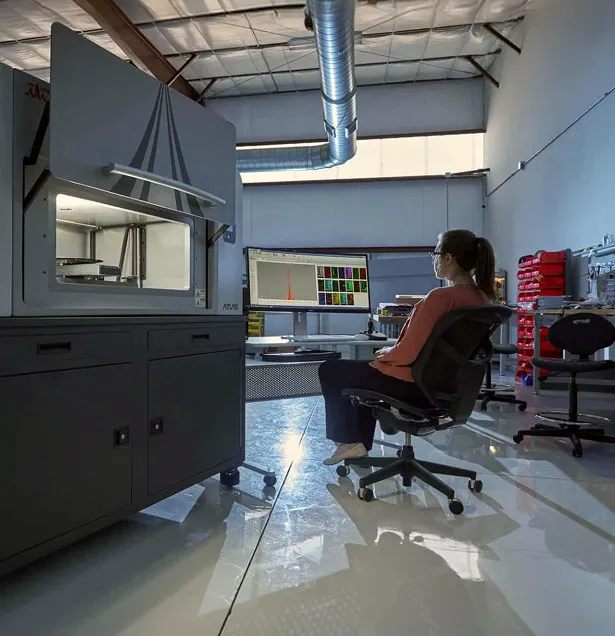
Match your elemental analysis technique to your sample size, feature scale, and analytical goals.
MicroXRF and scanning electron microscopy with energy-dispersive X-ray spectroscopy (SEM-EDS) both reveal how elements are distributed within a sample.
The key difference is scale: each technique supports different sample sizes, feature sizes, and analytical objectives.
SEM-EDS combines high-resolution microstructural imaging with elemental analysis of small features. MicroXRF maps elemental distributions across larger areas, often with substantially less sample preparation.
For many laboratories, choosing between them is less about which technique is better and more about which one fits the task.
Start with this question: Which technique is better suited to the scale and analytical objective of this sample?
How MicroXRF and SEM-EDS Work
MicroXRF: Elemental Mapping With a Focused X-Ray Beam
MicroXRF uses a focused X-ray beam to excite characteristic X-ray fluorescence from a sample.
As the beam scans the specimen, an energy-dispersive detector records a spectrum at each position.
The resulting elemental maps show compositional variation across small regions of interest or relatively large specimens.
Because MicroXRF uses X-rays rather than electrons for excitation, samples typically do not need to be electrically conductive and can often be examined at atmospheric pressure.
SEM-EDS: Microstructural Imaging With Elemental Analysis
SEM-EDS uses a focused electron beam to interact with the sample.
The microscope produces high-resolution electron images, while the EDS detector measures characteristic X-rays generated by the electron–sample interaction.
Together, these capabilities connect microstructural detail with elemental composition.
This makes SEM-EDS especially useful for investigating small features in their microstructural context.
Key Differences Between MicroXRF and SEM-EDS
Spatial Resolution
SEM-EDS typically offers finer spatial resolution than MicroXRF.
For nanoscale and sub-micron features, electron microscopy is usually the more appropriate tool.
MicroXRF works at a larger scale, although modern systems can use focused X-ray beams only a few micrometers in diameter.
This scale is well suited to features such as:
- Mineral grains
- Inclusions
- Coatings
- Reaction fronts
- Corrosion features
- Solder joints
- Contaminants
- Layered structures
Choose the spatial resolution that matches the feature you need to investigate.
Analysis Area
The area you need to examine can be as important as the size of an individual feature.
SEM-EDS supports detailed investigation of relatively small fields of view.
MicroXRF efficiently maps larger areas while preserving the broader compositional context.
This makes MicroXRF useful for examining an entire:
- Thin section
- Rock slab
- Electronic assembly
- Coating
- Biological specimen
- Cultural heritage object
- Industrial component
Use these larger-area maps to identify regions that merit higher-resolution investigation.
Sample Preparation
SEM analysis often requires more careful sample preparation.
Depending on the material and analytical objective, preparation may involve:
- Cutting
- Mounting
- Polishing
- Drying
- Conductive coating
- Vacuum-compatible preparation
MicroXRF often requires minimal preparation.
Many samples can be measured directly, especially when the goal is qualitative elemental mapping rather than high-accuracy quantitative analysis.
This can be valuable for fragile, valuable, irregular, or difficult-to-prepare materials.
Vacuum and Measurement Environment
Conventional SEM analysis takes place under vacuum.
This can limit analysis of hydrated specimens, volatile materials, large objects, and certain cultural heritage samples.
MicroXRF commonly operates at atmospheric pressure, with air, helium, or other measurement environments selected according to the elements of interest.
This flexibility simplifies measurement for many sample types.
Elemental Sensitivity
Both techniques detect a broad range of elements, but sensitivity varies with the sample and measurement conditions.
Key factors include:
- Element
- Matrix
- Detector configuration
- Acquisition conditions
- Sample geometry
- Excitation physics
MicroXRF often provides strong sensitivity for heavier elements, making it useful for locating trace- and minor-element features across relatively large areas.
SEM-EDS provides elemental information from small features at high spatial resolution.
Imaging and Microstructural Context
SEM produces detailed images of sample morphology and microstructure.
That imaging capability is a major advantage when interpreting the composition of individual features.
MicroXRF is primarily an elemental mapping technique, rather than a high-resolution morphological imaging method.
Combining optical images with XRF maps helps relate elemental distributions to visible sample features.
Applications for Each Technique
Typical MicroXRF Applications
MicroXRF is well suited to applications such as:
- Large-area elemental mapping
- Geological thin sections
- Critical-mineral exploration
- Coating and interface analysis
- Electronics
- Cultural heritage
- Corrosion studies
- Environmental samples
- Biological materials
Typical SEM-EDS Applications
SEM-EDS is well suited to applications such as:
- Fine microstructures
- Small particles
- Nanoscale or sub-micron features
- Fracture surfaces
- Microelectronics
- Detailed inclusion analysis
- High-resolution phase characterization
Use MicroXRF and SEM-EDS Together
For heterogeneous samples, MicroXRF and SEM-EDS often provide the most value as complementary tools.
Start with a large-area elemental survey, then investigate selected features at higher resolution:
- Use microXRF to map a large sample.
- Identify chemically unusual or important regions.
- Select targeted areas of interest.
- Transfer those regions to the SEM.
- Perform higher-resolution imaging and EDS analysis.
This workflow can reduce time spent searching in the SEM while retaining the sample’s larger compositional context.
Which Technique Is Right for Your Sample?
Choose MicroXRF for Larger-Area Elemental Mapping
Prioritize MicroXRF when your analytical goals include:
- Large analysis areas
- Minimal sample preparation
- Non-destructive analysis
- Trace-element mapping
- Atmospheric analysis
- Rapid survey of heterogeneous materials
Choose SEM-EDS for Fine Features and Microstructural Detail
Prioritize SEM-EDS when your analytical goals include:
- Maximum spatial resolution
- Microstructural imaging
- Very small features
- Correlation between morphology and composition
If your laboratory investigates heterogeneous materials across multiple length scales, consider how both techniques can support the workflow. MicroXRF and SEM-EDS are often complementary, not interchangeable.